Introduction to Liver MRI for Surgeons
- Diagnosis of Metastatic Liver Cancer -
Imaging investigations used in liver tumor diagnosis and liver MRI basics (2)
MR images of liver anatomy and surrounding organs
Images provided by: Hamamatsu University School of Medicine
Fig. 7 Axial images
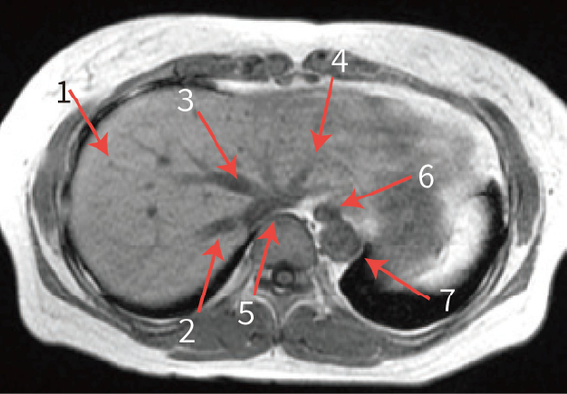
- 1.Liver
- 2.Right hepatic vein
- 3.Middle hepatic vein
- 4.Left hepatic vein
- 5.Inferior vena cava
- 6.Esophagus
- 7.Abdominal aorta
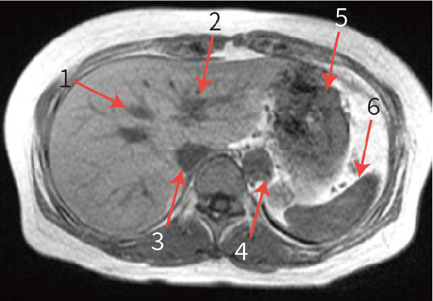
- 1.Middle hepatic vein
- 2.Umbilical portion
- 3.Inferior vena cava
- 4.Abdominal aorta
- 5.Stomach
- 6.Spleen
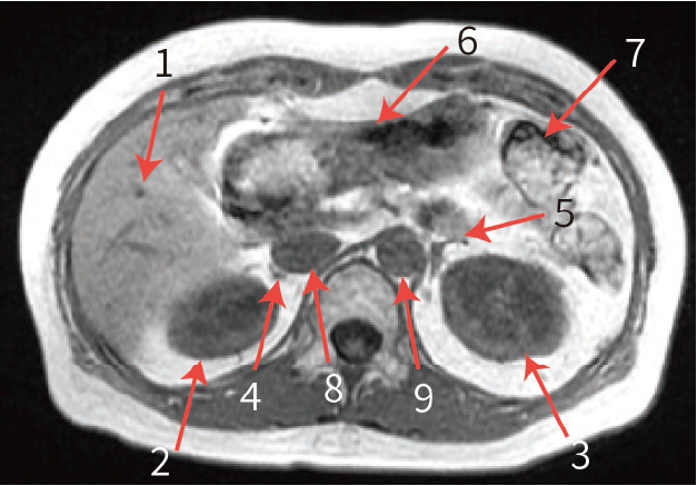
- 1.Liver
- 2.Right kidney
- 3.Left kidney
- 4.Right adrenal gland
- 5.Left adrenal gland
- 6.Stomach
- 7.Colon splenic flexure
- 8.Inferior vena cava
- 9.Abdominal aorta
Fig. 8 Coronal images
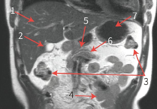
- 1.Liver
- 2.Gallbladder
- 3.Transverse colon
- 4.Jejunum
- 5.Pancreas
- 6.Main pancreatic duct
- 7.Stomach
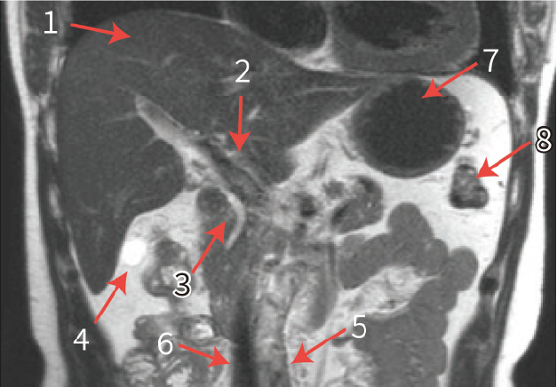
- 1.Liver
- 2.Portal vein
- 3.Common bile duct
- 4.Gallbladder
- 5.Abdominal aorta
- 6.Inferior vena cava
- 7.Stomach
- 8.Colon splenic flexure

- 1.Liver
- 2.Right adrenal gland
- 3.Right kidney
- 4.Left kidney
- 5.Spleen
- 6.Stomach
- 7.Descending colon
- 8.Major psoas muscle
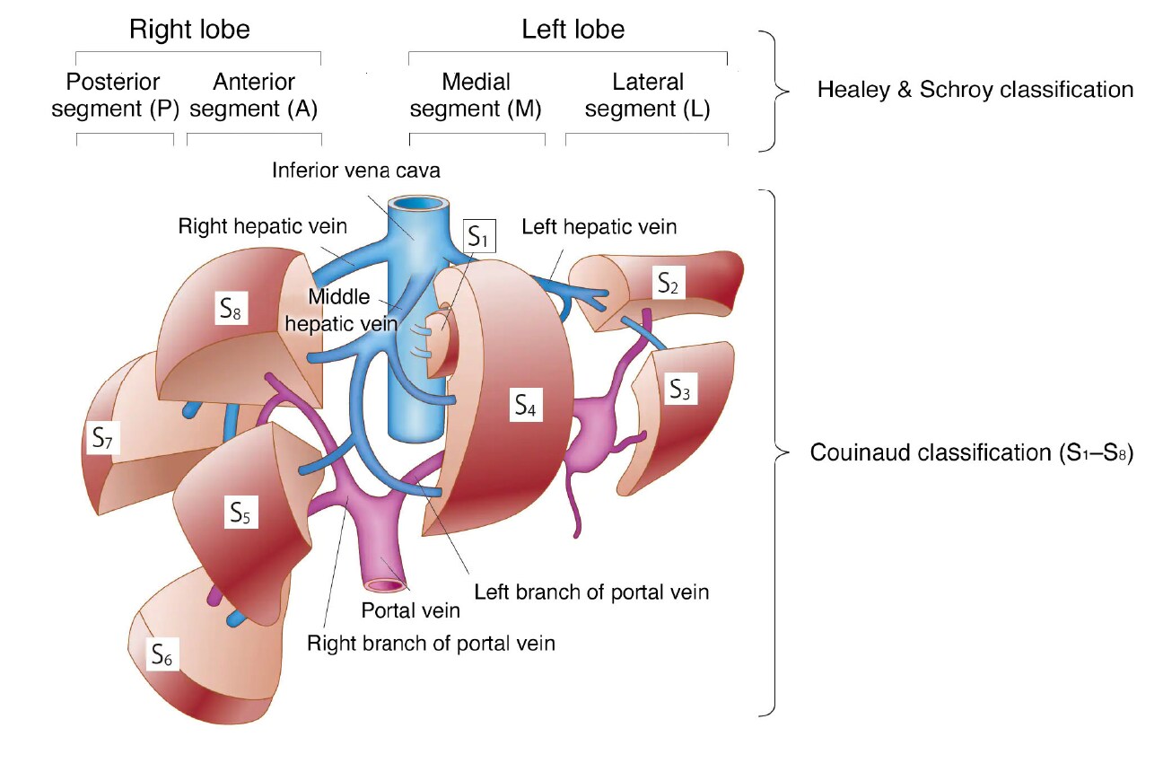
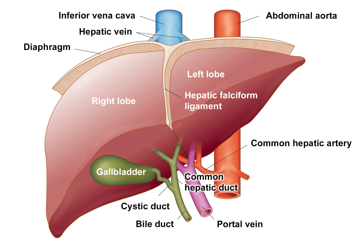
Fig. 9 Schematic view of the liver
MR images of liver segments
Images provided by: Hamamatsu University School of Medicine
Fig. 10 Hepatic segments in each cross-sectional image
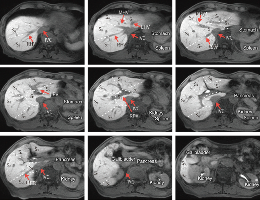
IRHV: inferior right hepatic vein
Fig. 11 Liver segments: Couinaud Classification and Healey & Schroy Classification