Optimising the arterial phase

Key Learnings:

  • Background of artifacts that can potentially impact the arterial phase of a Gadoxetate disodium(Gd-EOB-DTPA) Liver MRI
  • Theory of bolus geometry, relaxivity and timing the arterial phase in the liver with Gadoxetate disodium(Gd-EOB-DTPA)
  • Techniques to mitigate the appearance of artifacts in the arterial phase with Gadoxetate disodium(Gd-EOB-DTPA).

Kristie Smith is a qualified radiographer in Australia, topping her graduating class from University of Sydney in 2006. She has medical imaging experience in general x-ray, mammography, CT, fluoroscopy and over 10 years’ clinical experience in MRI; the last 6 years working as a Senior MR radiographer.

About this webinar:

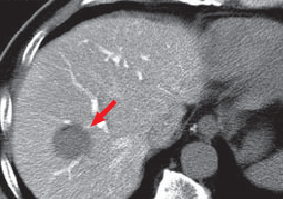
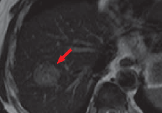
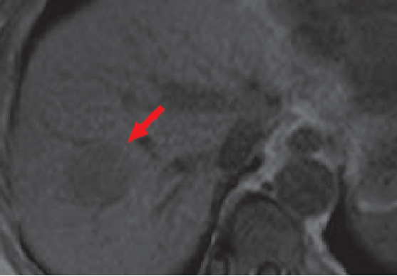
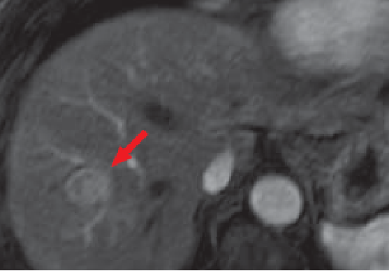
The arterial phase in a liver MRI with Gadoxetate disodium(Gd-EOB-DTPA) is important in identifying and classifying focal liver lesions based on their enhancement patterns. Transient respiratory motion and truncation artifact can potentially impact the image quality of the arterial phase. These artifacts can be potentially lessened through utilizing a number of strategies, including optimized Gadoxetate disodium(Gd-EOB-DTPA) injection, appropriate timing of the arterial phase and patient instructions, and using mutli-arterial sequences.

APAC.Liver360.video.Artifact.optimising the arterial phase 1/3

YYMMDD Author/Uploaded by

Lorem ipsum dolor sit amet, consectetur adipiscing elit, sed do eiusmod tempor incididunt ut labore et dolore magna aliqua.

Introduction and artifacts in liver imaging (10 minutes)

  • Hepatobiliary phase
  • Arterial phase & potential challenge (artifacts and timing)
  • Transient respiratory motion (TRM): definition, prevalence
  • Truncation artifact
  • Risk factor of TRM: Previous TRM, COPD, age, BMI, breath hold capacity

APAC.Liver360.video.Artifact.optimising the arterial phase 2/3

YYMMDD Author/Uploaded by

Lorem ipsum dolor sit amet, consectetur adipiscing elit, sed do eiusmod tempor incididunt ut labore et dolore magna aliqua.

Proposed strategies for arterial phase optimization: injection rate and acquisition timing (12.5 minutes)

  • Injection rate: slower injection rate for bolus, relaxivity, and protein binding
  • Power injection
  • Saline flush
  • Fluoroscopic triggering

APAC.Liver360.video.Artifact.optimising the arterial phase 3/3

YYMMDD Author/Uploaded by

Lorem ipsum dolor sit amet, consectetur adipiscing elit, sed do eiusmod tempor incididunt ut labore et dolore magna aliqua.

Proposed strategies for arterial phase optimization : Patient Preparation (10 minutes)

  • breath hold command
  • Sequence strategies: multi-arterial phases
  • Oxygen inhalation
  • Summary

About this webinar:

Patient’s background and MRI objectives

Patient’s background and MRI objectives

The patient was a male in his 60s, with chronic, non-B non-C hepatitis. At a different hospital, hepatitis was suspected, so the patient was hospitalized and ultrasonography was performed, showing a hepatic tumor. Contrast computed tomography (CT), and MRI using a nonspecific extracellular contrast agent, were then performed, but the tumor was difficult to distinguish, so the patient was referred to the authors’ hospital. After admission, angiographic CT was performed, followed by Primovist disodium gadoxetate contrast MRI 4 days later.

Legend: Angiographic CT

(a) CTAP 

Legend: Angiographic CT

CT under arterial portography [CTAP (a)] showed a perfusion defect approximately 35 mm in diameter in S8 of the liver (red arrow). In addition, in the early phase of CT during hepatic arteriography (CTHA; image not shown), dark staining was shown, and in the late phase a capsule-like structure was found.

(b) T2-weighted image

(b) T2-weighted image

(c) Pre-contrast T1-weighted image

(c) Pre-contrast T1-weighted image

MRI

The tumor (red arrow) appeared as a high-signal region in the T2-weighed image (b), and a low-signal region in the T1-weighted image (c).

(d) Arterial phase

(d) Arterial phase

(e) Hepatobiliary phase

(e) Hepatobiliary phase

Primovist disodium gadoxetate contrast MRI

The tumor (red arrow) appeared darkly stained in the arterial phase of Primovist disodium gadoxetate contrast MRI (d). During the hepatobiliary phase (e), 20 minutes after administration, most of the tumor showed the same signal as the liver parenchyma.

(f) Macroscopic observation

(f) Macroscopic observation

Pathology

The tumor was a green-brown color, reflecting biliary congestion. It was a hepatic tumor, showing moderate to high histopathological differentiation. The surrounding hepatic tissue had the appearance of chronic active hepatitis.

Impact of Primovist disodium gadoxetate on diagnosis

Tumor detectability in the hepatobiliary phase is high, and hepatocellular carcinoma (HCC) generally shows low signal intensity. However, tumors such as this case, with signal intensity similar to or higher than the hepatic parenchyma, also occur. With this case, macroscopic observation of an excised sample showed green coloration, indicating biliary congestion, and tumors with biliary congestion are termed “green hepatoma”.
Tumors showing similar signal intensity to the hepatic parenchyma may be overlooked with observation in the hepatobiliary phase alone. Green hepatoma can often be identified by T2-weighted imaging, dynamic MRI, etc., and is therefore detectible if a different sequence is followed. In the case of assessments using Primovist disodium gadoxetate, it is important to interpret images taking into consideration the existence of such tumors.

Impact of Primovist disodium gadoxetate on treatment

The present patient had a hepatic tumor that had developed in the context of chronic, non-B non-C hepatitis, and at the initial examination it was difficult to reach a definitive diagnosis as unifocal HCC. Contrast ultrasonography, CTHA/CTAP, and Primovist disodium gadoxetate contrast MRI were all performed, leading to a diagnosis of unifocal HCC, and surgical treatment was selected. With the abdominal contrast CT in current use, even when unifocal HCC is diagnosed, multiple tumors are often then diagnosed with CTHA/CTAP, but in the present case the finding was unifocal HCC with both Primovist disodium gadoxetate contrast MRI, and CTHA/CTAP. Furthermore, the HCC was confirmed to be unifocal by intraoperative ultrasonography.
The authors’ experience is that HCC with associated biliary congestion does not always have signal intensity similar to the hepatic parenchyma in the hepatobiliary phase of Primovist disodium gadoxetate contrast MRI, and may, as in the present case, have such similar signal intensity with associated low signal intensity, or may have high signal intensity. Preoperative diagnosis of biliary congestion is not linked to the treatment approach, but, as Primovist disodium gadoxetate contrast MRI generates nonspecific images, it is important to make the diagnosis by combining images from different phases rather than on the basis of the hepatobiliary phase alone.

  • *The case introduced is just one clinical case, so the results are not the same as for all cases.
  • *Please refer to the Package Insert for the effects and indications, dosage and administration method, and warnings, contraindications, and other precautions with use.