

The usefulness of EOB-MRI in differentiating focal nodular hyperplasia and subtype hepatocellular adenoma
Taipei Veterans General Hospital
Chien-An Liu, Division of Abdominal Imaging, Department of Radiology, Taipei Veterans General Hospital, Taipei, Taiwan.
Yi-Ping Hung, Department of Oncology, Taipei Veterans General Hospital, Taipei, Taiwan.
DATE : 2024
Introduction

Patient’s background
19 years old, Female, 48.0Kg
Disease(s): Hepatocellular adenoma (HCA)
Patient characteristics and purpose of contrast-enhanced MRI
This 19-year-old female had a past medical history of right adrenocortical carcinoma s/p surgical resection when she was two years old. She started to present with upper abdominal pain with a pain score of 8 out of 10. The pain is featured by the postprandial trigger, radiation to peri umbilicus, no specific exacerbating factor, and is able to be calmed with a painkiller. She visited a local hospital where the contrast-enhanced CT scan was done and showed a 10.0cm enhancing mass at the left lobe of the liver. Rather than metastatic lesions, the diagnosis favored focal nodular hyperplasia(FNH) or hepatocellular adenoma(HCA). Surgical resection was suggested, and she was referred to our institute. To achieve a more accurate diagnosis before surgery. The Gd-EOB-DTPA enhanced MRI was arranged for further evaluation.
Contrast agent used
Gadoxetate disodium(Gd-EOB-DTPA) injection, 0.1 mL/kg
Case description
In the EOB-MRI, the tumor revealed specific imaging characteristics (Figure 1.), and HCA with the IHCA subtype was impressed. She underwent lateral sectionectomy of the liver. Pathology reported steatosis, aggregation of inflammatory cells, and sinusoidal dilatation. Immunostain for beta-catenin didn’t show aberrant nuclear and cytoplasmic staining. Hence, the diagnosis of inflammatory hepatocellular adenoma (IHCA) without malignant transformation was made. Thereafter, she was regularly followed in our institute under stable conditions.
Imaging

CT exam shows right adrenalectomy with absence of right adrenal gland(A). Dynamic phase images demonstrate a 10cm mass at the lateral segment of the liver. There is no intratumoral hemorrhage, necrosis, or central scar(B). On contrast administration, the tumor demonstrates relatively homogeneous enhancement through arterial and portal venous phase images(C, D)without washout. The impression was FNH or HCA.
Fig. 1. Triple-phase liver CT
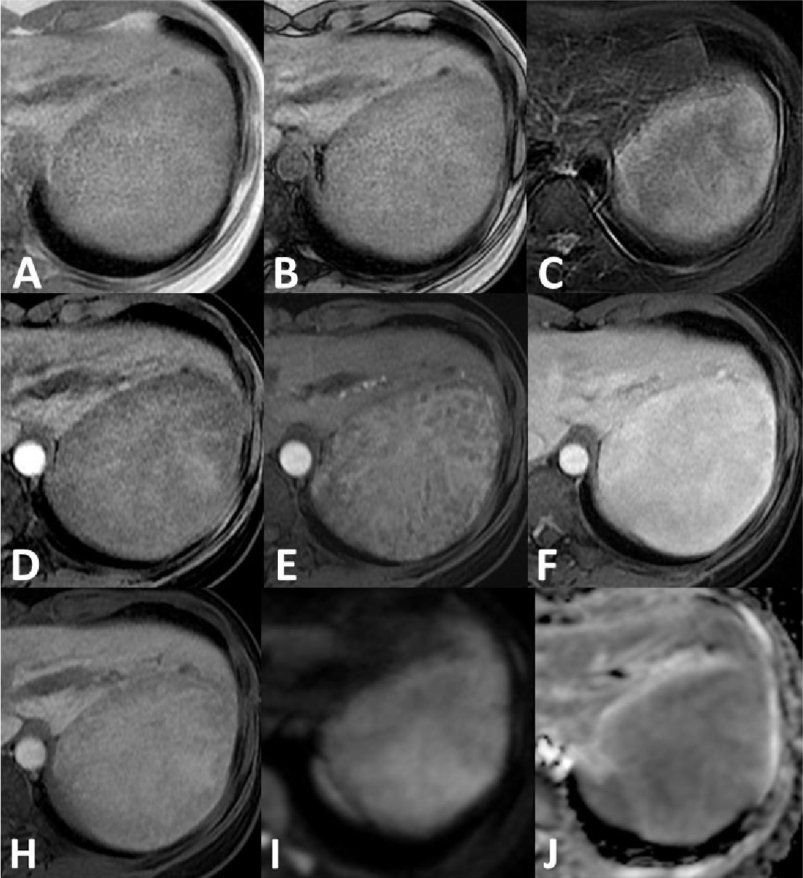
EOB-MRI before surgery shows some fat content of the tumor based on in- and out-of-phase images(A, B). (C)The tumor reveals high signal intensity in the fat-suppressed T2-weighted image, hypointensity in the precontrast T1-weighted image(D), heterogeneous enhancement in the arterial phase(E), and more homogenous enhancement in the portal phase(F). There is heterogeneous/patchy HBP Gd‑EOB uptake of the tumor(H), and it shows high signal intensity on diffusion-weighted image compared to the background liver(I) with low ADC value(J).
Fig. 2. Gd-EOB-DTPA enhanced MRI
Usefulness of contrast-enhanced MRI with Gadoxetate disodium(Gd-EOB-DTPA) in this case
Hepatocellular adenoma (HCA) is a rare benign tumor of the liver with unclear etiology. It is mainly found in young to middle-aged females and can be divided into four subtypes: HNF1A mutated adenoma(HHCA), inflammatory adenoma(IHCA), unclassified adenoma(UHCA), and β-catenin activated adenoma (bHCA). FNH and HCA both can show substantial enhancement(HHCA can present hypovascularity1) on contrast enhancement CT or gadolinium-enhanced MRI. FNH is commonly treated conservatively, while HCA should be resected because subtypes IHCA and b HCA have the tendency to undergo malignant transformation.
The presence of intralesional steatosis(most in HCA), lobulated margin(more in FNH) and central scar(more in FNH) can be used to differentiate FNH and HCA2. MRI features can be used to classify subtypes1,3,4. IHCA may appear hyperintense foci on T1 when bleeding and hyperintense rim of the lesion on T2w sequences, the so-called atoll sign3. In EOB-MRI, HCAs are generally hypointense during the hepatobiliary phase(HBP), but the β-catenin mutated subtype and up to a third of IHCAs are the exceptions to this characterization by showing hyperintense2,3.
In conclusion, the EOB-MRI provides a more accurate diagnosis than conventional triple-phase CT in this case and affects the treatment strategy. Furthermore, the imaging features of EOB-MRI added value for subtype differentiation of HCAs.
Imaging protocol
Devices used and contrast conditions
| MR device used | Signa HDxt 1.5T(GE Healthcare) |
| Autoinjector | LF |
| Workstation | ADW4.5 |
| Contrast enhancement condition | Dose(mL) | Infusion rate (mL/sec) | Imaging timing | |
| Gadoxetate disodium(Gd-EOB-DTPA) | 0.1mL/kg | 1mL/sec | ー | |
| Physiological saline chaser | 30ml | 1mL/sec |
| Imaging type | Imaging sequence | Imaging duration | TE (msec) | TR (msec) | FA (deg) | Fat Sat (Type) | NEX (Number of additions) | k-space | Slice thickness (mm) | FOV (mm) | Phase direction (Number of steps) | Lead direction (Number of matrices) | Slice gap (mm) | Number of slices |
| Dual echo | FSPGR (in & out of phase) | 20" | 2.2 | 178 | 80 | n | 1 | linear | 8 | 32 | 224 | 256 | 2 | 20 |
| With a contrast agent | ||||||||||||||
| Dynamic | LAVA (axial) | 20" | 3.1 (minumum) | 6.7 | 12 | y | 1 | linear | 3 | 32 | 192 | 288 | 0 | 100 |
| DWI | EPI (b=800sec/mm2) | 4'44" | 57.1 | 10000 | n | y | 4 | linear | 8 | 32 | 128 | 96 | 2 | 20 |
| T2WI | PROPELLER (axial) | 3' | 97 | 6000 | n | y | 1 | linear | 6 | 32 | 256 | 256 | 0 | 34 |
| HBP | LAVA (axial) | 20" | 3.1 (minimum) | 6.8 | 12 | y | 1 | linear | 3 | 32 | 192 | 288 | 0 | 100 |
References
BiseS, FrulioN, HocqueletA, AlbertiN, BlancJF, LaurentC, et al. New MRI features improve subtype classification of hepatocellular adenoma. Eur Radiol. 2019;29(5):2436-47.
AuerTA, Walter-RitteIT, GeiseID, SchöningW, SchmelzleM, MüllerT, et al. HBP-enhancing hepatocellular adenomas and how to discriminate them from FNH in Gd-EOB MRI, BMC Med Imaging [Internet]. 2021;21(1):1-11. Available from: https://doi.org/10.1186/s12880-021-00552-0
AuerTA, FehrenbachU, GrieserC, PenzkoferT, GeiselD, SchmelzleM, et al. Hepatocellular adenomas: is there additional value in using Gd-EOB-enhanced MRI for subtype differentiation? Eur Radiol. 2020:30(6):3497-506.
SchaibleJ, SchreyerAG, MehrabiA, Longericht, KieserM, KreimeyerS, et al. Subtyping of hepatocellular adenomas using Gd-EOB-DTPA: а qualitative and quantitative analysis. Acta radiol. 2023;64(7):2253-60.
- * The case introduced is just one clinical case, so the results are not the same as for all cases.
- * Please refer to the Package Insert for the effects and indications, dosage and administration method, and warnings, contraindications, and other precautions with use.
