

Hypervolemic hepatic mass
Kouseiren Takaoka Hospital
Dr. Yasushi Horichi, Dept. of Radiology
DATE : 2021
Introduction

Patient’s background
Female; 50s; body weight: 53.2 kg; focal nodular hyperplasia
Assessment objectives
On August 11, 2010, breast-conserving surgery was performed for left breast cancer.
After surgery, tamoxifen was administered orally and leuplin was administered by injection.
Simple computed tomography (CT) during monitoring, on September 8, 2016, suggested that a new space-occupying lesion had formed in S7 of the liver.
EOB-MRI was performed on October 3, 2016, as a thorough examination.
Contrast agent used
Gadoxetate disodium(Gd-EOB-DTPA) injection, 0.1 mL/kg
Case explanation
Simple CT during monitoring 6 years after surgery for left breast cancer showed an approximately circular, faint, low-absorption, space-occupying lesion with distinct borders that was 16 mm in diameter, in S7 of the liver, and it was suspected of being a metastatic hepatic tumor.
Hepatic EOB-MRI was performed as a thorough examination.
The possibility of the lesion being focal nodular hyperplasia was considered, but the high signal intensity with T2-weighted imaging was atypical of that, and the possibilities of it being metastatic liver cancer, hepatocellular carcinoma, or hepatocellular adenoma could not be ruled out.
A percutaneous needle biopsy was performed for confirmation, and focal nodular hyperplasia was diagnosed pathologically.
Imaging findings
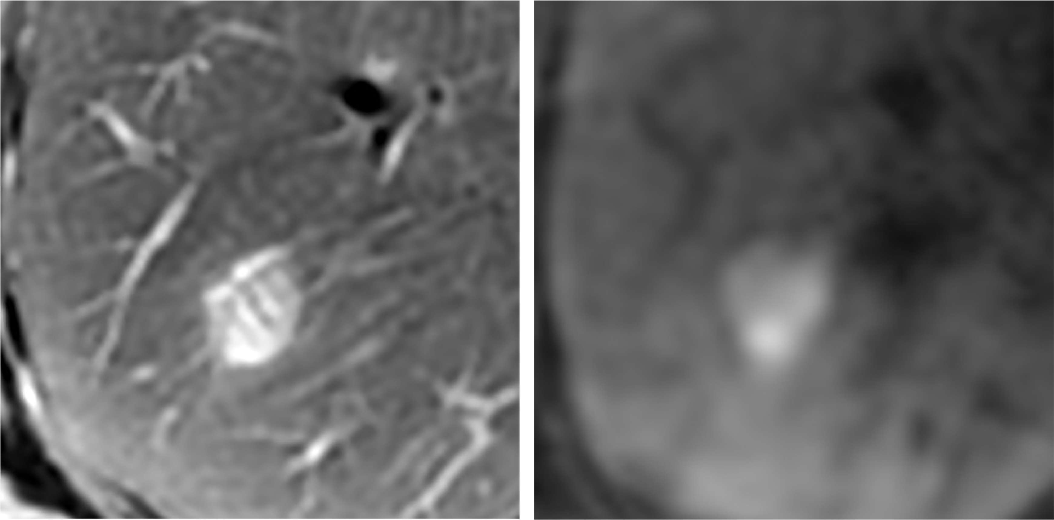
T2- and diffusion-weighted imaging showed the lesion to have distinct borders, and a high signal with a smooth margin.
T2-weighted imaging showed an internal septum- or band-shaped low signal, which may have been a central scar.
Fig. 1. T2- and diffusion-weighted imaging
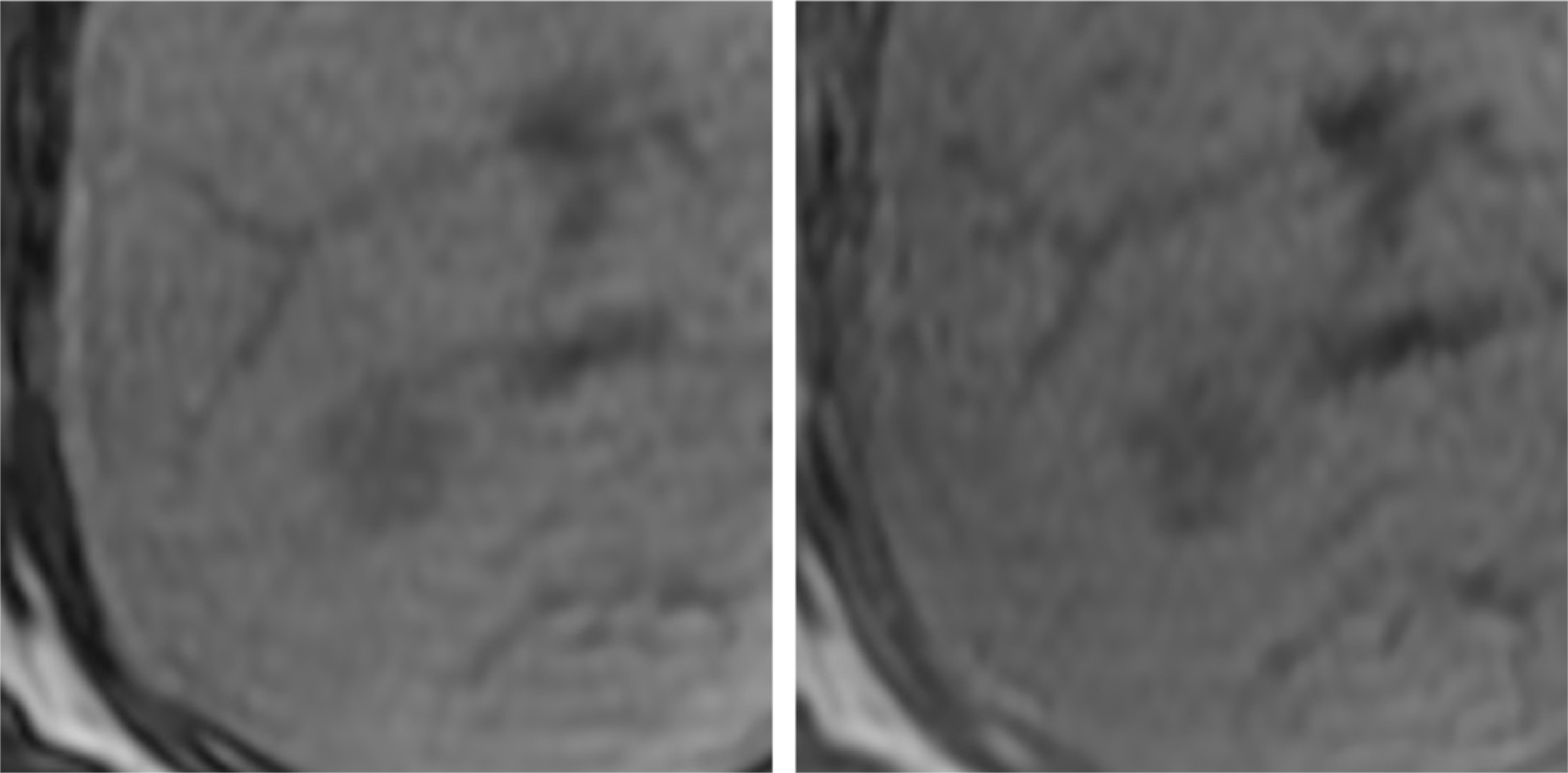
In-phase and opposed-phase T1-weighted imaging showed a low signal, and no clear fatty components were found in the lesion interior.
Fig. 2. T1-weighted imaging (in phase and opposed phase)
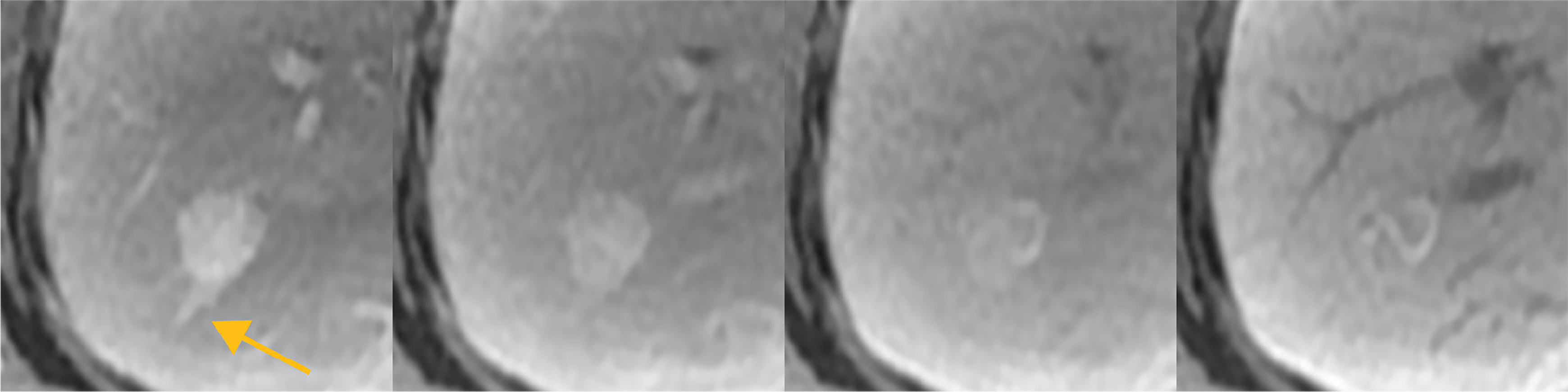
The early stage of dynamic imaging showed homogeneous dark staining.
Early-phase delineation of the hepatic vein was found on the peripheral side, and the possibility of it being an efferent vessel was considered.
In the late phase, wash-out was found in the central region, and in the transition phase an isosignal was shown in the center, and band-shaped dark staining was found to persist in the margins and interior.
Fig. 3. Dynamic imaging
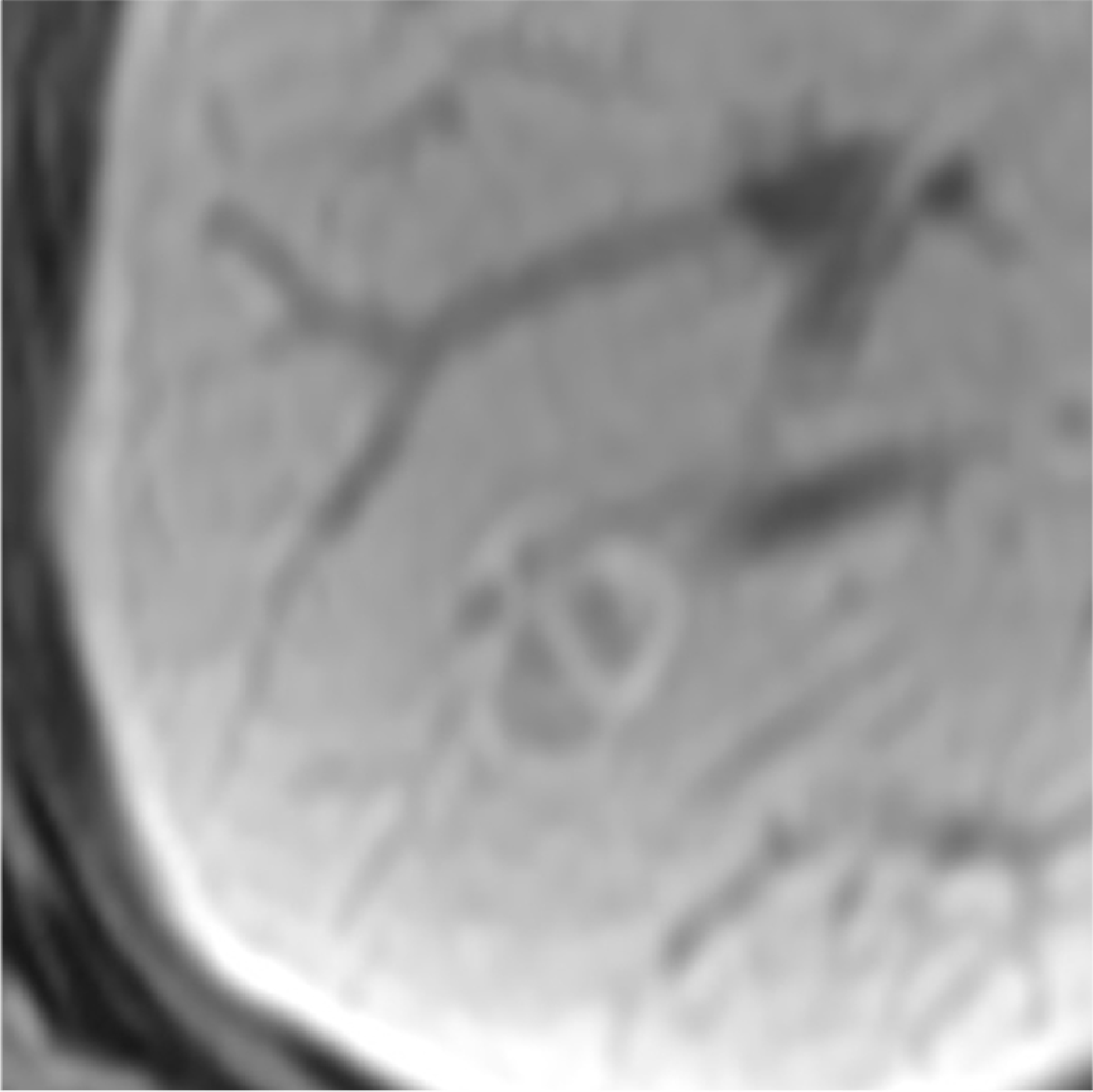
In the hepatobiliary phase, a band-shaped high signal was found in the margins and interior, similar to that found in the equilibrium phase of dynamic imaging.
Fig. 4. Hepatobiliary phase
Photography protocol
| Imaging type | Photography sequence | TE (msec) | TR (msec) | TI (msec) | FA (deg) | Fat sat (type) | ETL (number) | Holding breath (yes/no) |
| Dual echo | ー | ー | ー | ー | ー | ー | ー | ー |
| Contrast agent administration | ||||||||
| Dynamic | Dynamic SmartPrep | 1.5 | 3.0 | 1.5 | 12.0 | DIXON | ー | Yes |
| DWI | DWI Resp b1000 | 52.6 | 6315.8 | ー | 90 | ー | ー | No |
| T2WI | FS Resp PROPELLERA | 79.2 | 5714.3 | ー | 125 | chemi sat | 26 | No |
| HBP | LAVA-FLEX | 1.7 | 3.9 | ー | 12.0 | DIXON | ー | Yes |
| in phase | LAVA-FLEX | 2.2 | 3.9 | ー | 12.0 | ー | ー | Yes |
| out of phase | LAVA-FLEX | 1.1 | 3.9 | ー | 12.0 | ー | ー | Yes |
| Imaging type | NEX (calculation number) | Slice thickness (mm) | FOV (mm) | Phase direction (step number) | Read direction (matrix number) | Slice Gap (mm) | Slice number |
| Dual echo | ー | ー | ー | ー | ー | ー | ー |
| Contrast agent administration | |||||||
| Dynamic | 0.7 | 4 | 360 | 512 | 512 | 0 | 88 |
| DWI | 2.0 | 5 | 360 | 256 | 256 | 1 | 31 |
| T2WI | 1.5 | 5 | 360 | 512 | 512 | 1 | 31 |
| HBP | 0.7 | 4 | 360 | 512 | 512 | 0 | 92 |
| in phase | 0.7 | 4 | 360 | 512 | 512 | 0 | 92 |
| out of phase | 0.7 | 4 | 360 | 512 | 512 | 0 | 92 |
Devices used and contrast conditions
| MRI device | DISCOVERY MR750 |
| Automatic injection device | Sonic Shot GX (Nemoto Kyorindo) |
| Workstation | GE Advantage Workstation |
| Contrast conditions | Dose (mL) | Administration rate (mL/s) | Photography timing | |
| Gadoxetate disodium(Gd-EOB-DTPA) | 5.1 | 1 | smart prep. | |
| Physiological saline solution for flushing | 10 | 1 |
Usefulness of Gadoxetate disodium(Gd-EOB-DTPA) contrast MRI with this patient
Focal nodular hyperplasia is characterized by a fibrotic central scar, but small lesions are often difficult to identify, and sometimes difficult to distinguish from other hypervolemic tumors.
Whereas with metastatic liver cancer and hepatocellular carcinoma the efferent vessels are the surrounding sinusoids, with focal nodular hyperplasia they are the hepatic veins, and early-stage delineation of the surrounding hepatic veins is considered to be a relatively specific finding.
In approximately 40% of cases of focal nodular hyperplasia, donut-shaped dark staining at the margin is shown in the hepatobiliary phase, as with the present patient, and high OATP1B3 expression levels at the margin have also been reported.
EOB, combining characteristics of extracellular fluid contrast media and liver-specific contrast media, can be used for simultaneous evaluation of hemodynamics and hepatocyte function in lesions, and is an indispensable contrast medium for differentiation and diagnosis of hepatocytic nodules such as various types of hyperplasic nodule, hepatocellular adenoma, and hepatocellular carcinoma.
- *The case introduced is just one clinical case, so the results are not the same as for all cases.
- *Please refer to the Package Insert for the effects and indications, dosage and administration method, and warnings, contraindications, and other precautions with use.
