

Evaluation of cTACE and DEB-TACE therapeutic efficacy using Gadoxetate disodium(Gd-EOB-DTPA) contrast MRI (1)
Fukui-ken Saiseikai Hospital
Dept. of Diagnostic Radiology
Dr. Shiro Miyayama
DATE : 2021
Introduction
In Japan, the mainstream form of hepatic arterial chemoembolization (TACE) is conventional TACE (cTACE), in which lipiodol and a gelatin sponge (GS) are used. However, drug-eluting beads (DEBs) were introduced at the beginning of 2014. The basic principle of DEB-TACE is that blood vessels in and close to tumors are filled with granules that slowly release anticancer drugs, resulting in both ischemia and antitumor effects, and thus necrosis of the tumor. The therapeutic outcome has been evaluated as similar to that with cTACE, but DEB-TACE is expected to be effective for treating patients with advanced tumors and/or low hepatic functional reserve, and various studies have been performed at different facilities.
On the other hand, in the case of patients unresponsive to TACE, it is necessary to promptly decide upon the second-line therapeutic method, such as molecular-targeted drugs, and more accurate evaluation of therapeutic efficacy is therefore needed. Computed tomography (CT) and Gadoxetate disodium(Gd-EOB-DTPA) contrast MRI (EOB-MRI) are used to evaluate therapeutic effects after TACE, but evaluation after cTACE is sometimes hindered by the effects of lipiodol accumulated inside the tumor. In addition, it is sometimes difficult to distinguish minute residual tumors from treatment-related inflammation after DEB-TACE. In principle, the present author performs CT and EOB-MRI alternately at 2- to 3-month intervals after TACE, but evaluation by EOB-MRI is also performed if recurrence or persistence is suggested, but cannot be confirmed, by CT. Two patients with whom EOB-MRI was used for follow-up observation after TACE are presented below.
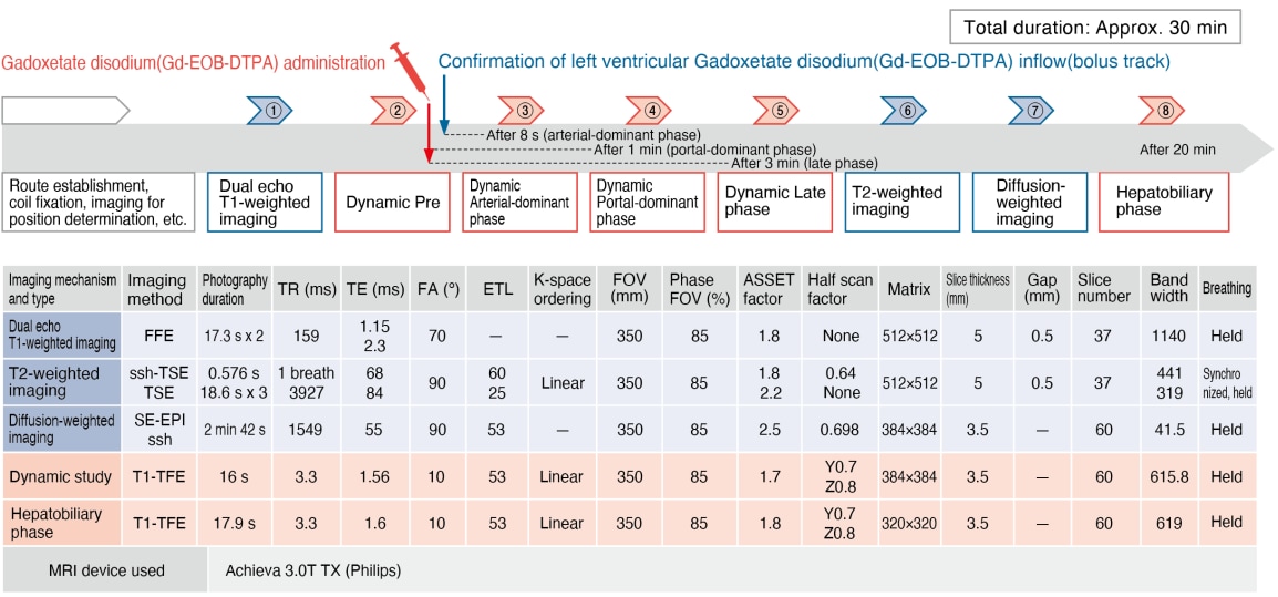
Magnetic resonance imaging (MRI) method using Gadoxetate disodium(Gd-EOB-DTPA)
Sequence and sequence parameters
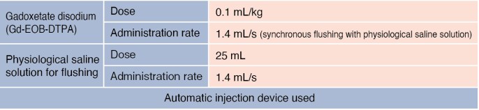
Contrast agent and administration method
CT method
Case presentation

Patient’s background and objectives of MRI
Male, 70s
Sudden and intense abdominal pain and blood pressure decrease occurred, and a local physician diagnosed intraperitoneal hemorrhage due to ruptured hepatocellular carcinoma. The patient was then transferred to the author’s hospital.
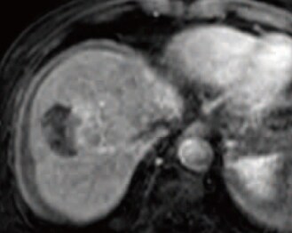
S8, lateral segment of left lobe: Arterial-dominant phase (a)
S4: Arterial-dominant phase (b)
Emergency digital subtraction angiography (DSA; c)
CT and DSA imaging
CT and DSA findings
The arterial-dominant phase of dynamic CT (a, b) showed three early-stage, dark-stained hepatic tumors, of which one was in S8, with a diameter of 5.5 cm; one in the lateral segment of the left lobe, with a diameter of 4.1 cm; and one (red arrow) in S4, with a diameter of 7 mm. Intraperitoneal hemorrhage and extravasation of contrast medium from the tumor in the lateral segment of the left lobe were also found (yellow arrow). The right-side part of the S8 tumor showed no early-stage dark staining.
Emergency DSA (c) showed vascular stenosis due to shock, and dark staining of the tumors in both lobes of the liver (blue arrows). Extravasation was found with imaging of the left hepatic artery, so embolization was performed using a GS, and hemostasis was achieved.
* The examination was performed using a 1.5T MRI device.
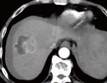
S8: Arterial-dominant phase (d)
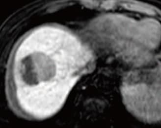
S8: Hepatobiliary phase (e)
S4: Hepatobiliary phase (f)
S8, S4: Simple CT (g)
EOB-MRI imaging*
EOB-MRI findings
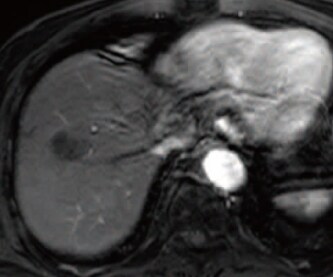
EOB-MRI performed after 8 days, with T1-weighting, showed, in the S8 tumor, a low signal when in phase, and a decreased signal in the right-side part of the tumor when out of phase, suggesting fat deposition. T2-weighted imaging showed a high signal (image not shown). In the arterial-dominant phase (d), the left part of the tumor showed early-stage dark staining, and in the hepatobiliary phase (e) the right-side part showed a distinct low signal, and the left part showed a slight low signal. In addition, the tumor in S4 showed a low signal in the hepatobiliary phase (f; red arrow), and intraperitoneal hemorrhage had decreased. After 18 days, cTACE was performed with the S8 and S4 tumors, using a suspension containing 8 mL of lipiodol, 30 mg of epirubicin, and 6 mg of mitomycin C, and a GS. Simple CT 1 week later (g) showed high-concentration accumulation of lipiodol in the tumor, except for the region with fat deposition.
S8: Arterial-dominant phase
(h)
EOB-MRI during the treatment course

S8: Arterial-dominant phase
(i)
EOB-MRI during the treatment course
S8: Arterial-dominant phase
(j)
CT during the treatment course
S8: Arterial-dominant phase
(k) *
EOB-MRI during the treatment course
EOB-MRI and CT findings
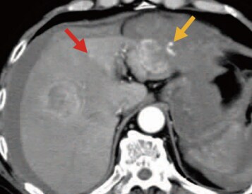
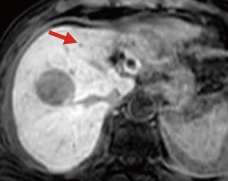
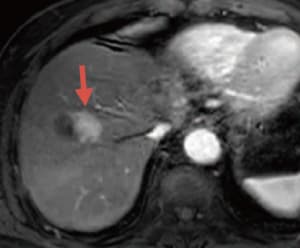
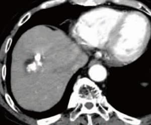
In the arterial-dominant phase of EOB-MRI after 9 months (h), tumor recurrence was found on the cranial side (red arrow), so cTACE was performed using a suspension of 4 mL of lipiodol, 20 mg of epirubicin, and 4 mg of mitomycin C, and a GS. However, in the arterial-dominant phase of EOB-MRI after 2 years and 10 months (i) tumor recurrence was found on the ventral side (yellow arrow), so cTACE was performed again using a suspension of 4 mL of lipiodol, 20 mg of epirubicin, and 4 mg of mitomycin C, and a GS. After 3 years and 7 months, no clear recurrence was found in the dynamic CT arterial-dominant phase (j), but in the EOB-MRI arterial-dominant phase (k) after 3 years and 10 months recurrence was found on the caudal side (blue arrows).

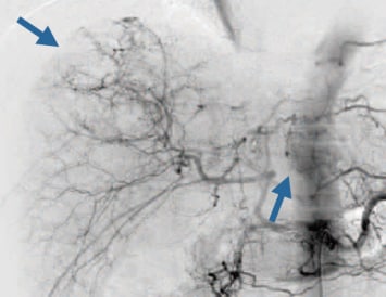
DSA (l)*
DSA imaging at the time of DEB-TACE
DSA findings
DEB-TACE was judged to be indicated because of local recurrence after cTACE. A faint tumor was found by DSA of the common hepatic artery (l; red arrows), and embolization of two A8 arteries (yellow arrows) was then performed using 0.2 mg of DEBs, prepared by diluting DC Beads (diameter: 100 to 300 μm) with an equal-volume mixture of Iopamirone 370 and physiological saline solution, and then impregnating 2 mg of the preparation with 50 mg of epirubicin.
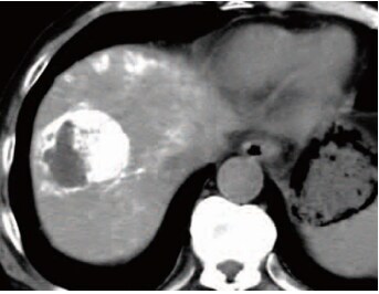
S8: Arterial-dominant phase (m)
CT imaging after DEB-TACE
S8: Arterial-dominant phase (n)*
EOB-MRI imaging after DEB-TACE
CT and EOB-MRI findings
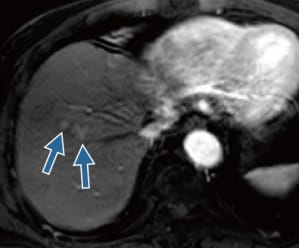
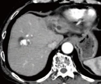
In the dynamic CT arterial-dominant phase (m) approximately 2 months after DEB-TACE, the presence of lipiodol made accurate judgment of therapeutic efficacy difficult, but in the EOB-MRI arterial-dominant phase (n) after 5 months (4 years and 4 months after the initial treatment), the early-stage dark staining found before treatment had disappeared, and there was judged to be no recurrence.
Usefulness of EOB-MRI diagnostic results with this patient
When judging therapeutic efficacy after cTACE, CT generated artifacts in the necrotic region of the tumor, due to concentrated lipiodol, which, together with the partial volume phenomenon, made accurate evaluation difficult, especially in the tumor marginal region. Therefore, there is a risk of the therapeutic efficacy being evaluated too highly, and it is preferable for evaluation to be performed with EOB-MRI. Even with the present patient, there may have been recurrence at the time of CT after 3 years and 7 months. In addition, even in the judgment about efficacy after DEB-TACE, EOB-MRI, which was not affected by previous lipiodol injection, clearly showed that there was no recurrence.
Precautions relating to administration
Administration to elderly patients
Elderly patients generally show depressed physiological function, and administration must be performed with care, and with sufficient monitoring of the patient’s condition.
- * The case introduced is just one clinical case, so the results are not the same as for all cases.
- * Please refer to the Package Insert for the effects and indications, dosage and administration method, and warnings, contraindications, and other precautions with use.
