

Hypervascular hepatocellular carcinoma not detected by computed tomography (CT)
Kyoto University, Graduate School of Medicine
Dept. of Diagnostic Imaging and Nuclear Medicine
Drs. Hiroyoshi Isoda, Kaori Togashi
DATE : 2021
Patient’s background

70s,
female.
The previous physician found hepatocellular carcinoma (HCC) in the left lobe of the liver by CT.
Investigation objectives
The objective was thorough preoperative assessment.
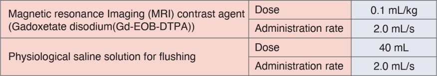
Contrast agent
Sequence and sequence parameters
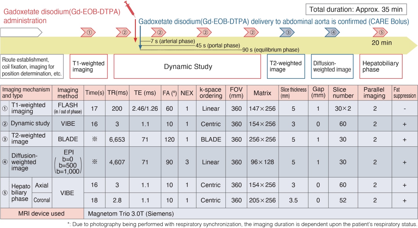
Findings
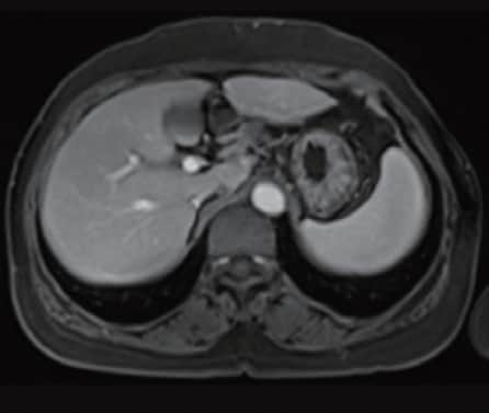
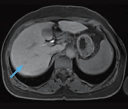
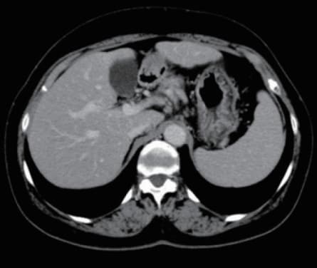
Lateral segment of left liver lobe
a) Arterial phase
b) Equilibrium phase
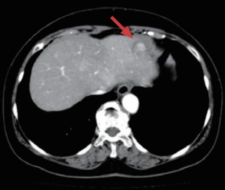
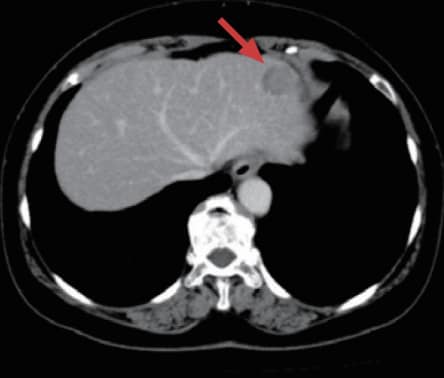
Multidetector CT
CT findings
A mass-type lesion (→), 19 mm in diameter, was found in the lateral segment of the left lobe of the liver, showing early-stage dark staining in the arterial phase (a), and distinct low absorption in the equilibrium phase (b).
c) Arterial phase
d) Hepatobiliary phase
Gadoxetate disodium(Gd-EOB-DTPA) contrast MRI
MRI findings
A mass-type lesion (→), 19 mm in diameter, was found in the lateral segment of the left lobe of the liver, showing early-stage dark staining in the arterial phase (c), and a distinct low signal in the hepatobiliary phase (d). Uptake of the contrast medium was observed in part of the tumor interior. These images suggest the presence of HCC.
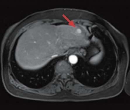
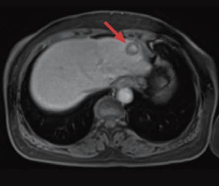
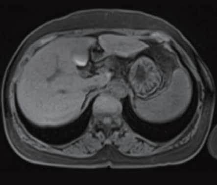
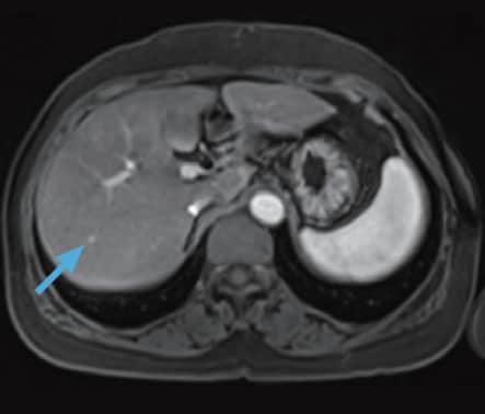
Posterior segment of right liver lobe
e) Pre-contrast, T1-weighted image
MRI
f) Equilibrium phase
g) Portal phase
Gadoxetate disodium(Gd-EOB-DTPA) contrast MRI
h) Hepatobiliary phase
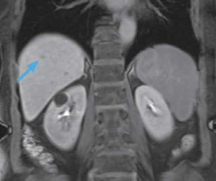
i) Hepatobiliary phase
Gadoxetate disodium(Gd-EOB-DTPA) contrast MRI
MRI findings
A small nodular lesion (→), 4 mm in diameter, was found in the posterior segment of the right lobe of the liver, and showed early-stage dark staining in the arterial phase (f). In pre-contrast, T1-weighted imaging (e) and the portal phase (g), this small nodule (→) could not be identified, but it showed a slightly low signal in the hepatobiliary phase (h, i). This nodule (→) was also suspected of being HCC.
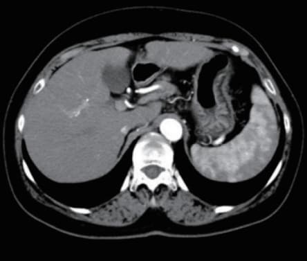
j) Early arterial phase

k) Late arterial phase
l) Equilibrium phase
MDCT
CT findings
HCC could not be identified in the early arterial phase (j), late arterial phase (k), or equilibrium phase (l) of dynamic CT.
Course of treatment
On the basis of preoperative diagnosis of HCC at two loci (→,→), partial resection was performed with the lesion (→) in the lateral segment of the left lobe of the liver, and intraoperative, ultrasound-guided radiofrequency ablation was performed with the lesion (→) in the posterior segment of the right lobe of the liver. HCC was diagnosed on the basis of the pathological findings.
Summary
As a result of recent hardware and software improvements, MRI image quality has improved markedly, and the clinical usefulness of MRI in the liver region has thus increased. As a result of the launch of 3T MRI, use of the better signal-to-noise ratio, which is the most important feature and advantage of 3T MRI, has enabled acquisition of images with high spatial resolution; and use of high tissue contrast, which is a feature of MRI, has enabled more accurate diagnosis of tumors.
Gadoxetate disodium(Gd-EOB-DTPA) contrast MRI is useful for detecting HCC and achieving a differential diagnosis, and the present authors have had experience with numerous patients, such as the present patient, with whom small HCC that could not be identified by dynamic CT was detectable by Gadoxetate disodium(Gd-EOB-DTPA) contrast MRI. For deciding upon the treatment plan for HCC, the importance of Gadoxetate disodium(Gd-EOB-DTPA) contrast MRI is expected to continue to increase.
Overview of adverse effects (total numbers in Japanese and overseas clinical studies at approval)
Of the total of 1,755 subjects, 76 (4.33%) suffered adverse effects. The principal adverse effects included the following: vasodilation (hot sensation, flushing): 16 patients (0.91%); nausea: 12 patients (0.68%); dysgeusia: 9 patients (0.51%); and headache: 8 patients (0.46%).
(total numbers in Japanese and overseas clinical studies at approval).
Dose and administration method
The usual adult dosage is 0.1 mL/kg, administered intravenously.
Precautions relating to administration
Administration to elderly patients
Elderly patients generally show depressed physiological function, so administration must be performed with care, and with sufficient monitoring of the patient’s condition.
- *The case introduced is just one clinical case, so the results are not the same as for all cases.
- *Please refer to the Package Insert for the effects and indications, dosage and administration method, and warnings, contraindications, and other precautions with use.
