

Benign lesion: Practical report (2)
Tottori University Hospital
Faculty of Medicine, Tottori University
Dept. of Pathophysiological and Therapeutic Science Division of Radiology:
Drs. Suguru Kakite, Shinya Fujii, Yasutoshi Ota, Yoshiko Kanasaki, Eiji Matsusue, Toshio Kaminou, Toshihide Ogawa
DATE : 2021
Diagnosis of hepatic hemangioma using 3-tesla magnetic resonance imaging (3T MRI)

Patient’s background and objectives of magnetic resonance imaging (MRI)
Female, 50s.
Abdominal ultrasonography was performed to monitor progression of cervical cancer, and showed a hepatic mass, so hemangioma was suspected, and computed tomography (CT) and MRI were performed.
This patient had typical hemangioma in S4 of the liver.
A noteworthy finding was the absence of dark staining in the late phase (f) of Gadoxetate disodium(Gd-EOB-DTPA) contrast MRI.
In the late phase, a hemangioma showing no dark staining was present, but contrast CT with this patient at the same time did show dark staining. This difference between MRI and CT is considered to be because uptake of Gadoxetate disodium(Gd-EOB-DTPA) by hepatocytes has already occurred by the late phase, making contrast enhancement in the hemangioma relatively indistinct.

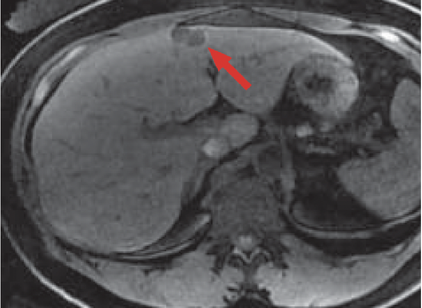
a) Pre-contrast

b) Post-contrast
Multidetector CT
Pre-contrast CT (a) showed low-absorption nodules in S4 of the liver (red arrow). Contrast CT (b) showed intense dark staining in S4 of the liver (red arrow). Imaging was performed 180 s after injection, at the same time as the late phase of Gadoxetate disodium(Gd-EOB-DTPA) contrast MRI.

c) T2-weighted imaging
d) Pre-contrast, T1-weighted imaging
MRI
Fat-suppressed, T2-weighted imaging (c) showed a lobular, high-signal mass in S4 (red arrow). Pre-contrast, T1-weighted imaging (d) showed a low-signal mass in S4 (red arrow).

e) Arterial phase

f) Portal phase

g) Late phase

h) Hepatobiliary phase (20 min after administration)
Gadoxetate disodium(Gd-EOB-DTPA) contrast MRI
In the arterial phase of Gadoxetate disodium(Gd-EOB-DTPA) contrast MRI (e), intense dark staining was found to the right side of the mass in S4 (red arrow).
In the portal phase (f), the mass in S4 showed increasing contrast enhancement from the margin to the interior (red arrow).
In the late phase (g), the mass in S4 showed a similar level of contrast to the surrounding hepatic tissues, and was indistinct (red arrow).
In the hepatobiliary phase (h) of Gadoxetate disodium(Gd-EOB-DTPA) contrast MRI, the mass in S4 gave a low signal, and no Gadoxetate disodium(Gd-EOB-DTPA) uptake was found (red arrow).
Summary
Gadoxetate disodium(Gd-EOB-DTPA) is a hepatocyte-specific contrast agent that is taken up by normal hepatocytes.
It is a derivative of conventional gadolinium contrast agents, and is used for T1-weighted imaging. It differs markedly from the superparamagnetic iron oxide preparations that are the liver-specific contrast agents in use to date, and use of 3-tesla liver acquisition with volume acceleration, or other three-dimensional gradient echo sequences with high sound/noise ratio, enables generation of high-resolution images that cannot be obtained using conventional liver-specific contrast agents.
In addition, in the initial phase of administration, Gadoxetate disodium(Gd-EOB-DTPA) is distributed similarly to the extracellular liquid contrast agent, and hemodynamic information is obtained by dynamic imaging. However, intracellular uptake by hepatocytes has been shown during the dynamic late phase, and the signal in the hepatic parenchyma increases. As a result, a mass such as a hemangioma that shows delayed dark staining may become more indistinct than when a conventional extracellular liquid contrast agent is used, and care is therefore essential in this respect.
- * The case introduced is just one clinical case, so the results are not the same as for all cases.
- * Please refer to the Package Insert for the effects and indications, dosage and administration method, and warnings, contraindications, and other precautions with use.
