

Practical report of metastatic hepatic cancer: Diagnosis of metastatic hepatic cancer using 3T MRI
Tottori University Hospital
Faculty of Medicine, Tottori University
Dept. of Pathophysiological and Therapeutic Science
Division of Radiology:
Drs. Suguru Kakite, Shinya Fujii, Yasutoshi Ota, Yoshiko Kanasaki, Eiji Matsusue, Toshio Kaminou, Toshihide Ogawa
DATE : 2021
Diagnosis of metastatic hepatic cancer using 3-tesla magnetic resonance imaging (3T MRI)
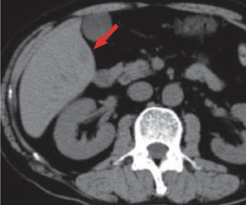
a) Pre-contrast
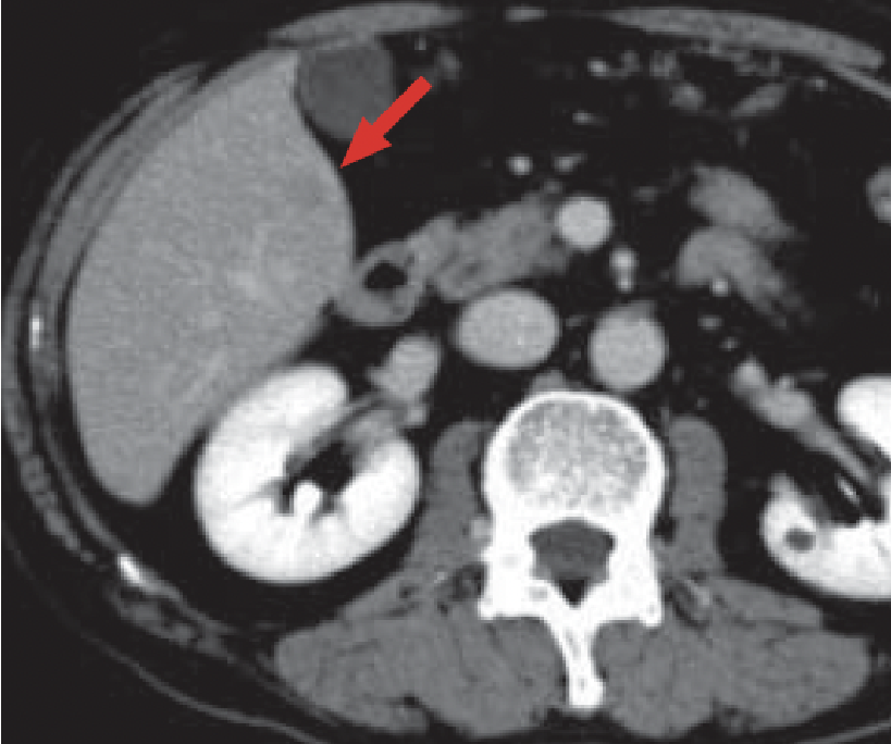
b) Post-contrast
Multidetector Computed Tomography (MDCT)
Pre-contrast CT (a) suggested the presence of a hepatic mass in S5 of the liver, showing faint low absorption, but it was indistinct (red arrow).
Contrast CT (b) suggested the presence of a faint, indistinct, low-absorption region in S5 of the liver, but this was not identifiable as a distinct mass (red arrow).
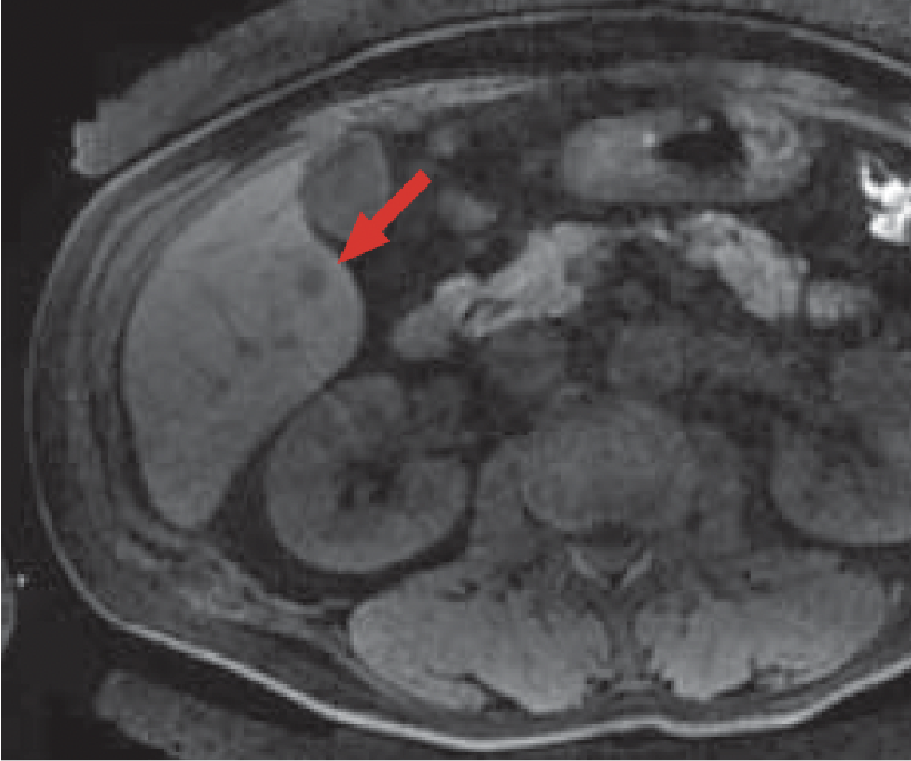
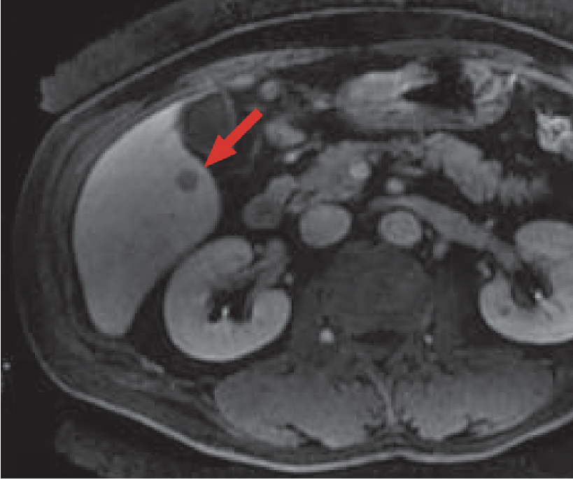
c) Pre-contrast, T1-weighted imaging
MRI
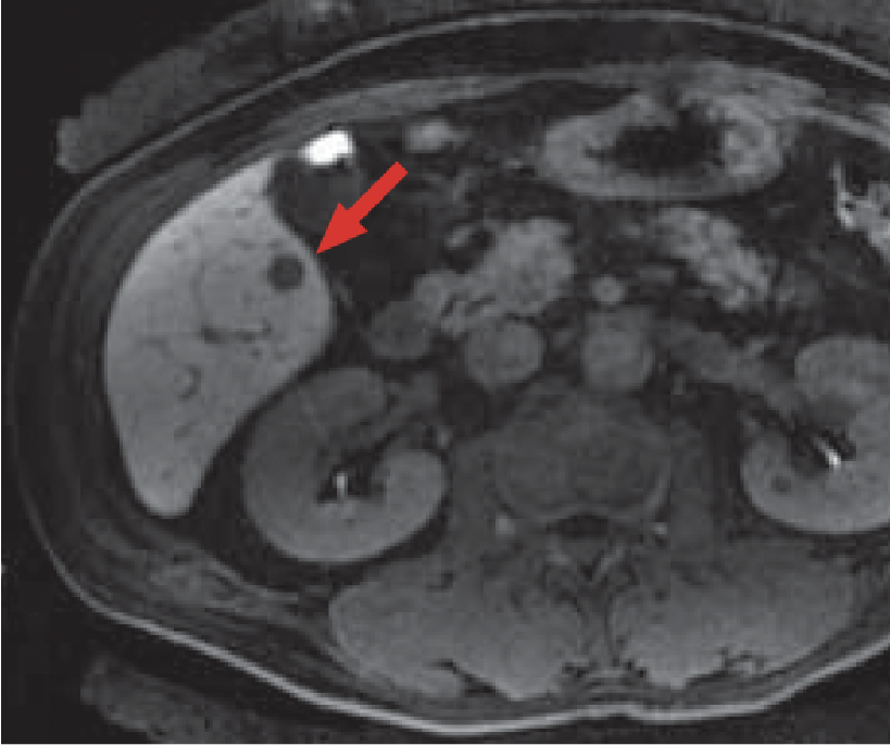
d) Arterial phase
e) Late phase
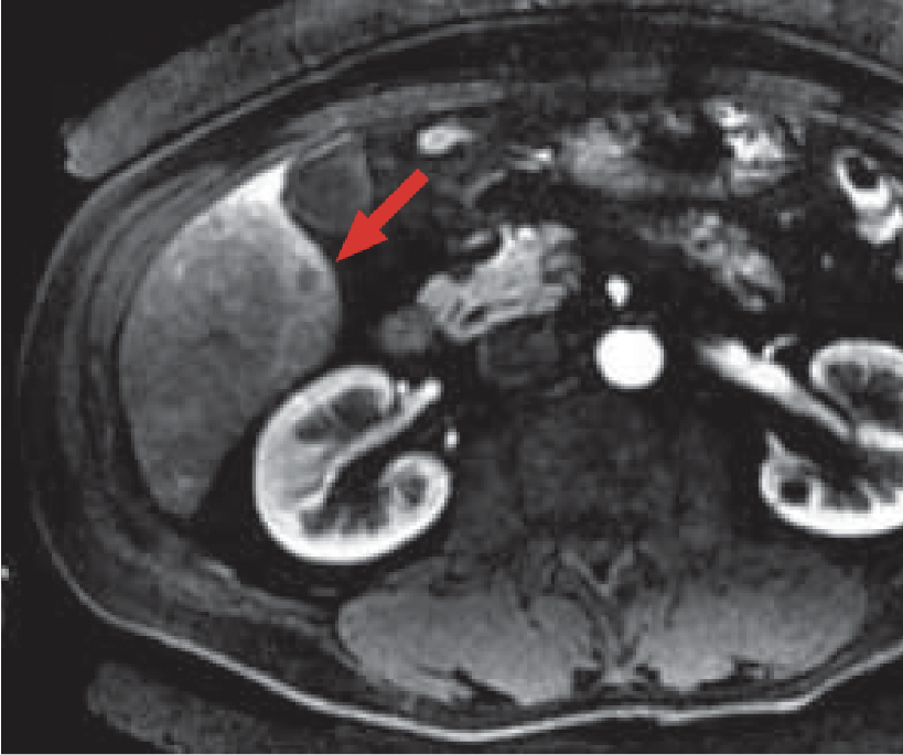
f) Hepatobiliary phase (20 minutes after administration)
Gadoxetate disodium(Gd-EOB-DTPA) contrast MRI
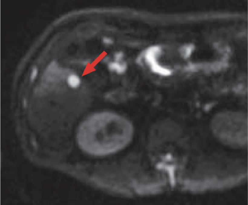
g) Diffusion-weighted imaging
MRI
In pre-contrast MRI (c), a mass giving a faint low signal was found in S5 of the liver.
In the Gadoxetate disodium(Gd-EOB-DTPA) contrast MRI arterial phase (d), the margin of the mass in S5 showed ring-shaped, faint contrast enhancement, and no clear contrast enhancement could be found in the central region of the mass (red arrow).
In the Gadoxetate disodium(Gd-EOB-DTPA) contrast MRI late phase (e), the mass in S5 gave a low signal (red arrow).
In the hepatobiliary phase (f) of Gadoxetate disodium(Gd-EOB-DTPA) contrast MRI, the mass in S5 was shown as a more distinct low-signal region than in the late phase (e; red arrow).
With diffusion-weighted imaging (g), the mass in S5 gave a high signal (red arrow).
Patient’s background and objectives of magnetic resonance imaging (MRI)
Male, 70s.
Gastric cancer was found with a gastric camera at a medical examination, so the patient was referred to and examined at Tottori University Hospital.
Contrast CT, performed as a search for metastases, suggested a mass in S5 of the liver, and MRI was performed for detailed examination approximately 1 week later.
Hepatic metastatic foci derived from advanced gastric cancer could not be confirmed by contrast CT (b), but were visualized distinctly by Gadoxetate disodium(Gd-EOB-DTPA) contrast MRI (d, e, f).
Hepatic metastases are usually shown as low-signal masses in the Gadoxetate disodium(Gd-EOB-DTPA) contrast MRI hepatobiliary phase (f). This was confirmed in the equilibrium phase (e), but contrast with the surrounding hepatic parenchyma was more distinct in the hepatobiliary phase (f). In addition, by performing dynamic MRI (d, e), it was possible to ascertain the hemodynamic status shown using conventional extracellular liquid contrast agents, enabling a qualitative diagnosis.
The administration volume of Gadoxetate disodium(Gd-EOB-DTPA) was half that of the gadolinium contrast media in use previously. Although there are concerns about reduction in contrast at the time of dynamic imaging, combination with 3-tesla liver acquisition with volume acceleration, which offers higher contrast resolution, enabled images to be generated to as high quality as dynamic images obtained using conventional extracellular liquid contrast agents.
Precautions relating to administration
Administration to elderly patients
Elderly patients generally show depressed physiological function, so administration must be performed with care, and with sufficient monitoring of the patient’s condition.
- *The case introduced is just one clinical case, so the results are not the same as for all cases.
- *Please refer to the Package Insert for the effects and indications, dosage and administration method, and warnings, contraindications, and other precautions with use.
