

Hyperintense HCC at hepatobiliary phase of EOB-MRI
Department of Medical Imaging and Intervention, Linkou Chang Gung Memorial Hospital. Taoyuan, Taiwan.
An-Hsin Chen
Department of Medical Imaging and Intervention, Linkou Chang Gung Memorial Hospital. Taoyuan, Taiwan.
Jeng-Hwei Tseng
Department of Medical Imaging and Intervention, Linkou Chang Gung Memorial Hospital. Taoyuan, Taiwan.
DATE : 2022
Patient characteristics

Patient’s background
68 years old, Male
Disease(s): Hepatocellular carcinoma (HCC)
Patient characteristics and purpose of contrast-enhanced MRI
This patient is a 68-year-old male with history of hepatitis B who had regular follow-up visits to GI clinic at local hospital, where a new 2-cm liver nodule was found. The patient was referred to our hospital for further MRI evaluation. He did not have any abnormalities in his laboratory test, including liver function tests and alpha-fetoprotein tumor marker.
Contrast agent used/ Dose
Gadoxetate disodium(Gd-EOB-DTPA) injection 0.1 ml/1kg (fix dose: 10 ml per patient)
Case description
EOB-MRI had revealed a 1.9-cm hyper-vascular nodule in S8 liver, with contrast medium pooling at hepatobiliary phase. Due to presence of capsule, HCC was firstly suspected rather than focal nodular hyperplasia (FNH). The patient was thus received S5/8 segmental hepatectomy and HCC (pT1) was confirmed by gross pathology.
Images
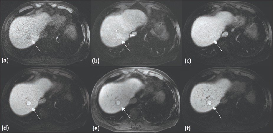
A 1.9-cm nodule in S8 liver (white arrow), appears hypointense on pre-contrast T1WI, with contrast pooling on hepatobiliary phase, which is classic characteristic for focal nodular hyperplasia (FNH). However, with presence of hypointense capsule at hepatobiliary phase, HCC is concerned.
- Figure 1. EOB-MRI T1-weighted image (T1WI), pre- and post-contrast dynamic phase
(a) T1 pre-contrast; (b) T1 post-contrast arterial phase ;
(c) T1 post-contrast venous phase; (d) T1 post-contrast 5 mins
(e) T1 post-contrast 10 mins; (f) T1 post-contrast 20 mins
(a) T1 pre-contrast; (b) T1 post-contrast arterial phase ;
(c) T1 post-contrast venous phase; (d) T1 post-contrast 5 mins
(e) T1 post-contrast 10 mins; (f) T1 post-contrast 20 mins
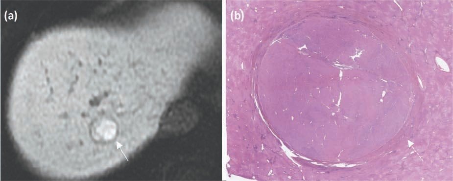
The hypointense capsule on hepatobiliary phase of EOB-MRI (white arrow) is well depicted on the microscopic pathologic section (dashed arrow).
- Figure 2. (a) T1 post-contrast 20 mins, focused on the S8 lesion
(b) Microscopic image of the lesion
(b) Microscopic image of the lesion
Usefulness of contrast-enhanced MRI with Gadoxetate disodium(Gd-EOB-DTPA) in this case
According to Fujita et al. (Radiographics. 2020 Jan-Feb;40(1):72-94.), about 10-15% of HCCs are hyperintense during hepatobiliary phase of EOB-MRI, due to preserved OATP1B3, which is an uptake transporter of gadoxetic acid and usually has stronger expression in FNH. FNH usually consists of peripheral hyperplastic hepatocytes and central scars; and the areas of hyperplastic hepatocytes would appear ring or donut-like hyperintensity at hepatobiliary phase. In terms of our case here, the presence of capsule, non-ring or non-donut like enhancement on hepatobiliary phase would provide some clue for diagnosing HCC.
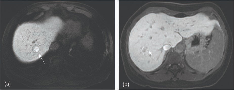
Presence of capsule with rather homogenous hyperintensity at hepatobiliary phase in HCC (white arrow) is different from FNH (dashed line), which would not have tumor capsule, and its peripheral hyperplastic hepatocytes would uptake the gadoxetic acid resulting in ring or donut-like enhancement at hepatobiliary phase.
- Figure 3. Different hyperintense appearance between HCC and FNH
(a) The aforementioned case, T1 post-contrast, hepatobiliary phase
(b) Another patient with FNH, T1 post-contrast hepatobiliary phase
(a) The aforementioned case, T1 post-contrast, hepatobiliary phase
(b) Another patient with FNH, T1 post-contrast hepatobiliary phase
Imaging protocol
Devices used
| MR device used | GE Signa HDxt |
|---|---|
| Autoinjector | n/a |
| Workstation | AW VolumeShare 5 |
| Contrast enhancement condition | Dose(mL) | Infusion rate (mL/sec) | Imaging timing | |
|---|---|---|---|---|
| Gadoxetate disodium(Gd-EOB-DTPA) | 10 ml | 1 ml/s | 40 mins | |
| Physiological saline chaser | 10 ml | 1 ml/s |
| Imaging type | Imaging sequence | Imaging duration | TE (msec) | TR (msec) | TI (msec) | FA (deg) | Fat Sat (Type) | ETL (Number) | Breath hold (Present/Absent) | NEX (Number of additions) | Slice thickness (mm) | FOV (mm) | Rectangular FOV (%) | Lead direction (Number of matrices) | Slice gap (mm) | Number of slices |
|---|---|---|---|---|---|---|---|---|---|---|---|---|---|---|---|---|
| Dynamic-C(-) | LAVA | 20s | 1.7 | 3.9 | 7 | 15 | special | 1 | present | 0.73 | 4.0 | 40 | 80 | 320x192 | 0 | 96 |
| With a contrast agent | ||||||||||||||||
| Dynamic-C(+) A | LAVA | 20s | 1.7 | 3.9 | 7 | 15 | special | 1 | present | 0.73 | 4.0 | 40 | 80 | 320x192 | 0 | 96 |
| Dynamic-C(+) V | LAVA | 20s | 1.7 | 3.9 | 7 | 15 | special | 1 | absent | 0.73 | 4.0 | 40 | 80 | 320x192 | 0 | 96 |
| Dynamic-C(+) 5mins | LAVA | 20s | 1.7 | 3.9 | 7 | 15 | special | 1 | present | 0.73 | 4.0 | 40 | 80 | 320x192 | 0 | 96 |
| Dynamic-C(+) 10mins | LAVA | 20s | 1.7 | 3.9 | 7 | 15 | special | 1 | present | 0.73 | 4.0 | 40 | 80 | 320x192 | 0 | 96 |
| Dynamic-C(+) 20mins | LAVA | 20s | 1.7 | 3.9 | 7 | 15 | special | 1 | present | 0.73 | 4.0 | 40 | 80 | 320x192 | 0 | 96 |
- *The case introduced is just one clinical case, so the results are not the same as for all cases.
- *Please refer to the Package Insert for the effects and indications, dosage and administration method, and warnings, contraindications, and other precautions with use.
