

Benign lesion: Practical report (1)
University of Tsukuba
Graduate School of Comprehensive Human Sciences
Majors of Medical Sciences
Dept. of Diagnostic Radiology
Drs. Katsuhiro Nasu and Manabu Minami
DATE : 2021
Focal nodular hyperplasia (FNH)

Patient’s background and objectives of MRI
Female, 40s. A hypervascular tumor was found in the posterior segment of the liver. No definitive diagnosis was made, but the patient was monitored for 8 years. No clear increase in tumor size was found during that time. Although no pathological diagnosis was made, FNH was suspected on the basis of the time-course. The patient was allergic to the iodine contrast agent, so follow-up was by simple MRI.
During the follow-up period, superparamagnetic iron oxide (SPIO) contrast MRI and a dynamic study using Gd-DTPA were performed (photographs not shown).

- a) Pre-contrast T2-weighted image
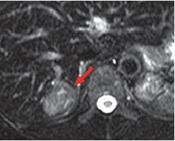
- b) SPIO contrast T2-weighted image
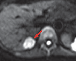
- c) Diffusion-weighted image
- d) Pre-contrast T1-weighted image
MRI
Comparison of T2-weighted images before (a) and after (b) SPIO contrast showed no clear SPIO uptake by the tumor (red arrow).
The tumor showed a distinct high signal even with diffusion-weighted imaging (c), and it showed a heterogeneous low signal with pre-contrast, T1-weighted imaging (d; red arrow).
This finding was nonspecific, and FNH could not be diagnosed definitively on this basis alone.
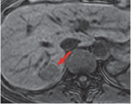
- e) Arterial phase
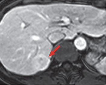
- f) Portal phase
Gadoxetate disodium(Gd-EOB-DTPA) contrast MRI
The tumor showed intense contrast enhancement in the arterial phase (e), and for 180 s after contrast agent administration in the portal phase (f) it showed a similar signal to the surrounding hepatic tissue (red arrow).
Up to this time, it had not been possible to verify the central scar in the tumor or blood vessels passing through the tumor center.
In a dynamic study using Gd-DTPA, performed during the follow-up period, the findings were similar to those detailed above (photographs not shown).
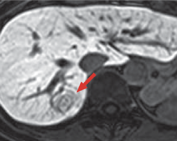
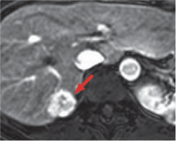
- g) Hepatobiliary phase

- h) Minimal intensity projection
Gadoxetate disodium(Gd-EOB-DTPA) contrast MRI
In the hepatobiliary phase (g), homogeneous uptake of Gadoxetate disodium(Gd-EOB-DTPA) by the tumor parenchyma was demonstrated, although it did not reach the normal hepatic parenchyma. In addition, blood vessels passing through the tumor interior were imaged distinctly, and it was confirmed that they were continuous from the hepatic portal region (red arrow). An essential point to note is that the contrast enhancement inside the blood vessels had almost disappeared by this time. Blood vessels shown in the hepatobiliary phase (g) could be observed distinctly when a minimal-intensity projection image (h) was prepared (blue arrow).
There is room for debate as to whether these blood vessels were arteries, but, taking the clinical time-course into consideration, it seems reasonable to consider that the imaging was of abnormal blood vessels passing through the FNH interior.
Information about diagnosis of FNH obtained by Gadoxetate disodium(Gd-EOB-DTPA) contrast magnetic resonance imaging (MRI) with this patient
As Gadoxetate disodium(Gd-EOB-DTPA) is taken up by hepatocytes immediately after administration, the blood concentration decreases at a speed that could not be imagined with the contrast media in previous use.
Therefore, the concept of the equilibrium phase, as relating to conventional extracellular contrast media, might not be applicable to this contrast medium. This property can be either advantageous or disadvantageous, but at least for FNH diagnosis it is probably a major advantage. In the hepatobiliary phase of Gadoxetate disodium(Gd-EOB-DTPA) contrast MRI, it can be shown on the basis of Gadoxetate disodium(Gd-EOB-DTPA) uptake by the tumor that the tumor is of hepatocyte origin. In addition, on the basis of Gadoxetate disodium(Gd-EOB-DTPA) blood concentration decrease, abnormal blood vessels passing through the tumor interior can be readily recognized. Furthermore, one of the properties of this tumor, hypervascularity, can be demonstrated on the basis of the high-image-quality arterial phase. In summary, it is possible to obtain all information needed for a diagnosis of FNH by Gadoxetate disodium(Gd-EOB-DTPA) contrast MRI.
- *The case introduced is just one clinical case, so the results are not the same as for all cases.
- *Please refer to the Package Insert for the effects and indications, dosage and administration method, and warnings, contraindications, and other precautions with use.
