

Mass-forming intrahepatic cholangiocarcinoma: Pathology and comparison
Tokyo Women's Medical University
Dr. Shun-ichi Ariizumi
DATE : 2009
Mass-forming intrahepatic cholangiocarcinoma

Patient’s background and objectives of MRI
Male, 41 years old.
Primary complaint: Right hypochondrial pain
History of current condition: In 2009, the patient was examined at a local hospital for the primary complaint of right hypochondrial pain.
A tumor 8 cm in diameter was found in the right lobe of the liver, and the patient was therefore referred to the author’s hospital.
Blood chemistry test results: Leukocytes: 6400 /μL; hemoglobin: 12 g/dL; platelets: 35 × 104 /μL; AST: 29 IU/L; ALT: 71 IU/L; total bilirubin: 0.2 mg/dL; albumin: 3.5 g/dL; ALP: 635 IU/L; γGTP: 124 IU/L; prothrombin time: 94%; ICGR15: 5%; Child-Pugh class: A; HBs antigen: negative; HCV antibody: negative; CEA: 2.4 ng/mL; and CA19-9: 420 U/mL.
Abdominal CT findings
A segmented tumor 7.5 cm in diameter was found in the right lobe of the liver. The arterial phase of contrast computed tomography (CT) showed the tumor to have low absorption, and even in the delayed phase it had low absorption (photograph not shown), but part of the interior showed delayed dark staining. In addition, an intrahepatic metastasis 1.5 cm in diameter was found in the peritumoral region.
EOB-MRI findings
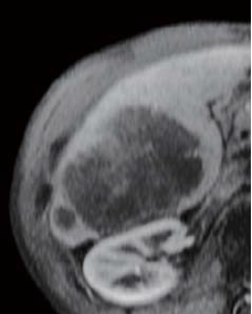
In-phase, T1-weighted imaging showed a low signal inside the tumor, and T2-weighted imaging showed a high signal (photographs not shown). Pre-contrast imaging gave a low signal, and in the dynamic study there was a low signal 20 s after administration, and 60 and 120 s after administration there was a low signal, but also delayed dark staining in the tumor interior. Even in the hepatobiliary phase, a low signal with associated delayed dark staining was seen in the tumor interior.
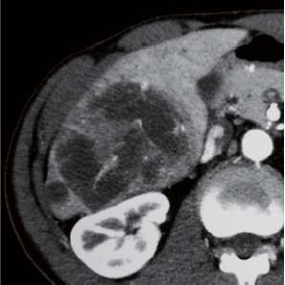
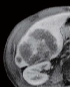
Contrast CT arterial phase
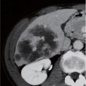
Contrast CT equilibrium phase
Abdominal CT findings
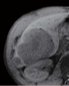
Pre-contrast
EOB-MRI findings
20 s after administration
60 s after administration
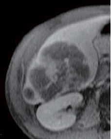
120 s after administration
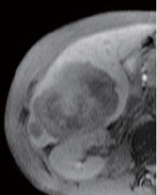
Hepatobiliary phase
EOB-MRI findings
Surgery
Right hepatectomy and lymph node dissection were performed.
Macroscopic observation of resection samples
A white, hard, lobulated mass without capsule formation was found, so the cancer was of the mass-forming type. There was a small intrahepatic metastasis by the side of the above.
Histopathology findings
(i) The principal nodule and satellite nodules are shown. The principal nodule has irregular, dark-colored regions scattered on the central side. (ii) “a” shows that atypical epithelial cells form glandular lumens, or in some cases the lumens are indistinct and have a cord-like form. Interstitial fibrous tissue can be seen between the above.
The cancer is an adenocarcinoma with low to moderate differentiation. (iii) “b” indicates a region that has undergone fibrosis, with atrophy of cancer cells, and voids left after loss of cancer cells.
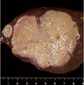
Findings with macroscopic
observation
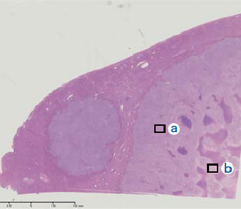
(i)
Histopathology findings
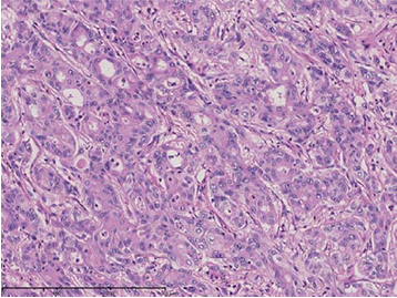
(ii) Magnification of “a”
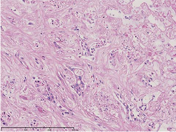
(iii) Magnification of “b”
Histopathology findings
- *The case introduced is just one clinical case, so the results are not the same as for all cases.
- *Please refer to the Package Insert for the effects and indications, dosage and administration method, and warnings, contraindications, and other precautions with use.
