

Hepatic metastases of colorectal cancer: Pathology and comparison
Tokyo Women's Medical University
Dr. Shun-ichi Ariizumi
DATE : 2009
Metastatic liver cancer

Patient’s background and objectives of MRI
Male, 77 years old.
Primary complaint: None
History of current condition: In 2007, the patient was diagnosed as having type-3 ascending colon cancer with synchronous hepatic metastases, and right hemicolectomy and right hepatectomy were performed. The histopathological diagnosis was highly differentiated adenocarcinoma, SE, with no lymph node metastases, and a total of five hepatic metastases up to 6 cm in diameter. The patient subsequently underwent chemotherapy at a local hospital, but in 2008 hepatic metastasis was found in the remaining liver, and he was again referred.
Blood chemistry test results: Leukocytes: 4570 /μL; hemoglobin: 15.5 g/dL; platelets: 17 × 104 /μL; AST: 34 IU/L; ALT: 32 IU/L; total bilirubin: 0.6 mg/dL; albumin: 4.0 g/dL; prothrombin time: 89%; ICGR15: 28%; Child-Pugh class: A; HBs antigen: negative; HCV antibody: negative; CEA: 54 ng/mL; and CA19-9: 155 U/mL.
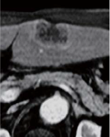
Abdominal CT findings
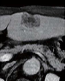
A tumor 4 cm in diameter, with an irregular margin, was found in S3 of the lateral segment of the liver. The tumor in S3 was shown by simple CT to have low absorption, and in the contrast CT equilibrium phase it was shown to have low absorption with contrast enhancement in the marginal region. The liver surface was slightly concave, and cancer navel formation was suspected.
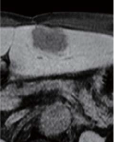
EOB-MRI findings
The tumor interior had a low signal in in-phase, T1-weighted imaging, and a high signal in T2-weighted imaging (photographs not shown). Pre-contrast, the tumor had a low signal, and in the dynamic study it had a low signal 20 s after administration, and 60 and 120 s after administration the margins showed mild dark staining, whereas the interior showed a low signal. The liver surface was concave, as in the CT findings. Even in the hepatobiliary phase, there was a distinct low signal.
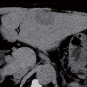
Simple CT
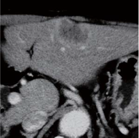
Contrast CT equilibrium phase
Abdominal CT findings
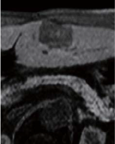
Pre-contrast
EOB-MRI findings
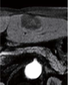
20 s after administration
60 s after administration
120 s after administration
Hepatobiliary phase
EOB-MRI findings
Surgery
Partial S3 resection was performed.
Macroscopic observation of resection samples
The tumor was a white color, with a cancer navel formed on the liver surface.
Histopathology findings
(i) The center of the tumor had a large necrotic region, and the marginal area had a region with dense proliferation of cancer cells. (ii) “a” indicates a region with numerous cells in the margin, and atypical columnar cells forming the lumen, the image showing moderately differentiated adenocarcinoma. (iii) “b” indicates a large necrotic region in the center, and insular fibrous tissue is present, surrounded by viable cancer cells.
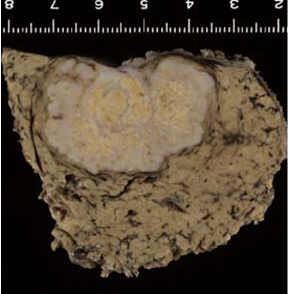
Findings with macroscopic
observation
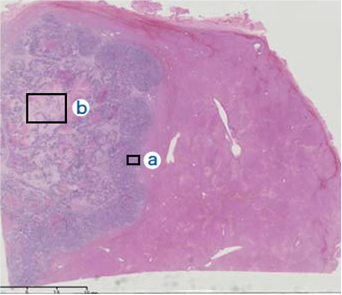
(i)
Histopathology findings
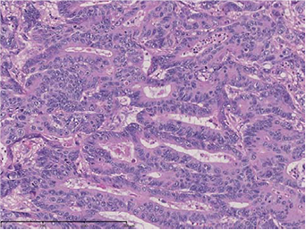
(ii) Magnification of “a”
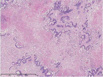
(iii) Magnification of “b”
Histopathology findings
Precautions for use
Administration to elderly patients
Elderly patients generally show depressed physiological function, and administration must therefore be performed with care, and with sufficient monitoring of the patient’s condition.
- *The case introduced is just one clinical case, so the results are not the same as for all cases.
- *Please refer to the Package Insert for the effects and indications, dosage and administration method, and warnings, contraindications, and other precautions with use.
