

Early-stage hepatocellular carcinoma (HCC): Pathology and comparison
Tokyo Women's Medical University Hospital
Dr. Shun-ichi Ariizumi
DATE : 2009
Small-nodular HCC with indistinct margins (early-stage HCC)
HCC in posterior segment
Abdominal CT findings
The tumor in the posterior segment (S6) was shown as a minor, low-absorption region in the equilibrium phase of contrast computed tomography (CT).
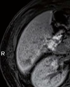
EOB-MRI findings
A minor, low-signal region was present before contrast, but in the dynamic study it was shown with a signal approximately equal to that in the circumferential liver, and was indistinct.
However, it was recognized as a distinct, low-signal region 1 cm in diameter in the hepatobiliary phase.
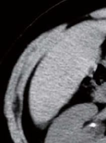
Simple CT
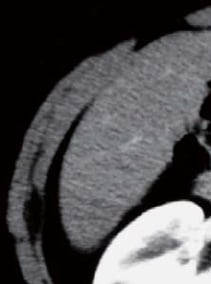
Contrast CT arterial phase
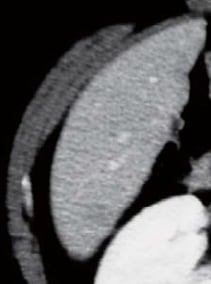
Contrast CT equilibrium phase
Abdominal CT findings
Pre-contrast
EOB-MRI findings
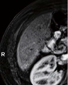
20 s after administration
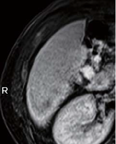
60 s after administration
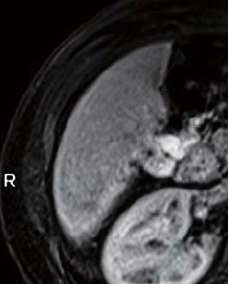
120 s after administration
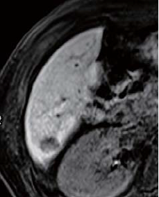
Hepatobiliary phase
EOB-MRI findings
Surgery
The S6 tumor was not clearly shown even by intraoperative ultrasonography, but part of it underwent colorization when an indocyanine green (ICG), infra-red camera system (Photo Dynamic Eye [PDE]) was used, and that part was excised.
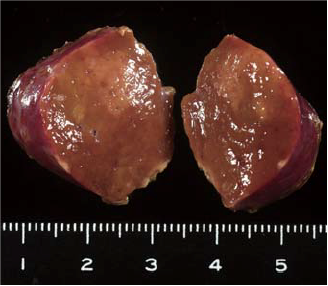
Macroscopic observation of resection samples
The S6 tumor was of a small-nodular type, with indistinct margins, and was a yellow-brown region with coloration differing only slightly from the non-cancerous hepatic tissue.
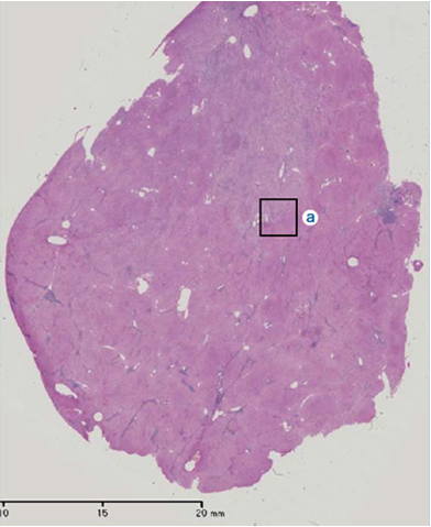
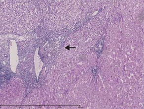
Histopathology findings
(i) Loupe image of a hematoxylin-eosin-stained specimen of the indistinct-margin nodule, at the left cut surface where macroscopic findings were made. A region was shown in which the existence or otherwise of the portal region below the hepatic capsule was unclear. “a” indicates the portal region in the marginal part of early-stage HCC. (ii) The upper, left part is early-stage HCC, the lower, right part is non-cancerous hepatic tissue, and between those is the portal region. (iii) Observation with Victoria blue staining clearly showed portal region infiltration (arrow).
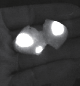
ICG infra-red camera system
Findings with macroscopic observation
(i)
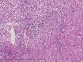
(ii) Magnification of “a”
(iii) Magnification of “a” (stained with Victoria blue)
Histopathology findings
- *The case introduced is just one clinical case, so the results are not the same as for all cases.
- *Please refer to the Package Insert for the effects and indications, dosage and administration method, and warnings, contraindications, and other precautions with use.
