

Nodule-in-nodule hepatocellular carcinoma (HCC): Pathology and comparison
Tokyo Women's Medical University Hospital
Dr. Shun-ichi Ariizumi
DATE : 2009
Nodule-in-nodule HCC

Male, 59 years old.
Primary complaint: None
History of current condition: In July 2008, hepatic dysfunction was found in a health check-up, and abdominal computed tomography (CT) was performed, suggesting HCC.
Hematology and blood chemistry test results: Leukocyte count: 5,820 /µL; hemoglobin: 16 g/dL; platelet count: 9.2 × 10⁴ /µL; aspartate aminotransferase: 30 IU/L; alanine aminotransferase: 46 IU/L; total bilirubin: 0.4 mg/dL; albumin: 4.6 g/dL; ICGR15: 7%; HBs antigen: positive; HBe antigen: negative; HBe antibody: positive; HCV antibody: negative; alpha-fetoprotein: 3 ng/mL; and PIVKA-II: 100 mAU/mL.
HCC in anterior segment
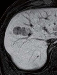
Abdominal CT findings
The tumor in the anterior segment had approximately equal absorption in simple CT, whereas with contrast CT a high-absorption region 1 cm in diameter was found in the arterial phase, and a faint low-absorption region 2 cm in diameter was found in the equilibrium phase
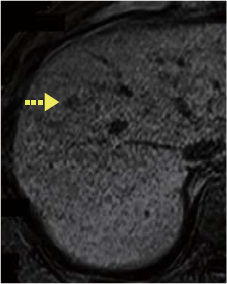
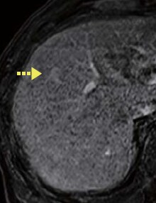
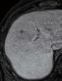
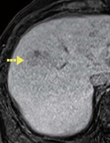
EOB-MRI findings
The pre-contrast image showed a somewhat low-signal region 1 cm in diameter. In the dynamic study, this tumor showed a somewhat high signal 20 s after administration, and a low signal 120 s after administration. However, in the hepatobiliary phase, in the area surrounding this tumor a low-signal region with distinct margins, 2 cm in diameter, was found, and a low-signal region consisting of two masses was also found, linked to the tumor on the medial side (the S6 lesion is shown on p. 6).
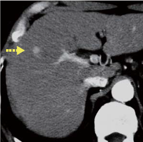
Contrast CT arterial phase
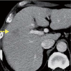
Contrast CT equilibrium phase
Abdominal CT findings
Pre-contrast
EOB-MRI findings
20 s after
administration
60 s after
administration
120 s after
administration
Hepatobiliary phase
EOB-MRI findings
Surgery
Anterior segment resection and partial S6 resection were performed.
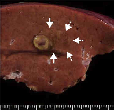
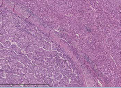
Macroscopic observation of resection samples
The tumors in the anterior segment were one that was 1 cm in diameter, with a swollen sectional surface, and one that was 2 cm in diameter, had indistinct margins, and was a somewhat different color from the surrounding non-cancer hepatic tissues (the S6 findings are shown on p. 7).
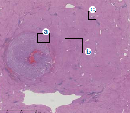
Histopathology findings
- (1)The nodular region with indistinct margins, to the right side of the simple nodule, was a region of early-stage HCC, and “a” shows the capsule of the simple nodule.
- (2)Inside the capsule there was HCC with a thick, moderately differentiated, cord-like structure, and the capsule exterior also had a structure similar to the portal region, initially appearing like non-cancer tissue.
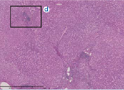
- (3)The portal area in the central part of the region “b” is shown with “d”.
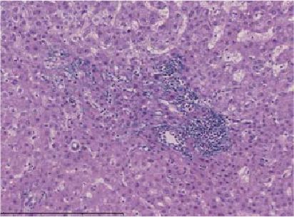
- (4)With magnification of the area shown by “d”, the image shows hepatocytes between the elastic fibers, which are stained with Victoria blue.
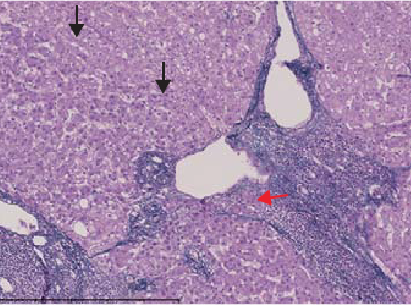
- (5)Magnification of the area shown by “c” shows infiltration (red arrow) into the portal region of the marginal region (black arrow) of the early-stage HCC.
Findings
with macroscopic observation
Histopathology findings (1)
(2) Magnification of “a”
(3) Magnification of “b”
(4) Magnification of “d”
(stained with Victoria blue)
(5) Magnification of “c”
(stained with Victoria blue)
Histopathology findings
- *The case introduced is just one clinical case, so the results are not the same as for all cases.
- *Please refer to the Package Insert for the effects and indications, dosage and administration method, and warnings, contraindications, and other precautions with use.
